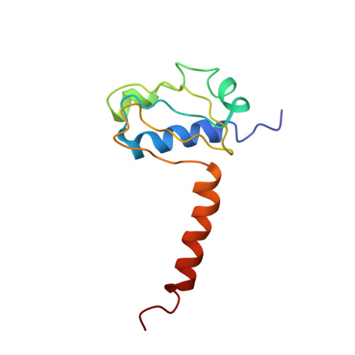
3LG8

Crystal structure of the C-terminal part of subunit E (E101-206) from Methanocaldococcus jannaschii of A1AO ATP synthase


Experimental Data Snapshot

  • Method: X-RAY DIFFRACTION
  • Resolution: 4.10 Å
  • R-Value Free: 0.381 
  • R-Value Work: 0.353 
  • R-Value Observed: 0.356 

wwPDB Validation   3D Report Full Report


This is version 1.4 of the entry. See complete history


Literature

Crystal and solution structure of the C-terminal part of the Methanocaldococcus jannaschii A1AO ATP synthase subunit E revealed by X-ray diffraction and small-angle X-ray scattering

Balakrishna, A.M.Manimekalai, M.S.S.Hunke, C.Gayen, S.Rossle, M.Jeyakanthan, J.Gruber, G.

(2010) J Bioenerg Biomembr 42: 311-320

  • DOI: https://doi.org/10.1007/s10863-010-9298-3
  • Primary Citation of Related Structures:  
    3LG8

  • PubMed Abstract: 

    The structure of the C-terminus of subunit E (E(101-206)) of Methanocaldococcus jannaschii A-ATP synthase was determined at 4.1 A. E(101-206) consist of a N-terminal globular domain with three alpha-helices and four antiparallel beta-strands and an alpha-helix at the very C-terminus. Comparison of M. jannaschii E(101-206) with the C-terminus E(81-198) subunit E from Pyrococcus horikoshii OT3 revealed that the kink in the C-terminal alpha-helix of E(81-198), involved in dimer formation, is absent in M. jannaschii E(101-206). Whereas a major dimeric surface interface is present between the P. horikoshii E(81-198) molecules in the asymmetric unit, no such interaction could be found in the M. jannaschii E(101-206) molecules. To verify the oligomeric behaviour, the low resolution structure of the recombinant E(85-206) from M. jannaschii was determined using small angle X-ray scattering. Rigid body modeling of two copies of one of the monomer established a fit with a tail to tail arrangement.


  • Organizational Affiliation

    School of Biological Sciences, Nanyang Technological University, Singapore, Republic of Singapore.


Macromolecules
Find similar proteins by:  (by identity cutoff)  |  3D Structure
Entity ID: 1
MoleculeChains Sequence LengthOrganismDetailsImage
A-type ATP synthase subunit E
A, B
106Methanocaldococcus jannaschiiMutation(s): 0 
Gene Names: atpEMJ0220
UniProt
Find proteins for Q57673 (Methanocaldococcus jannaschii (strain ATCC 43067 / DSM 2661 / JAL-1 / JCM 10045 / NBRC 100440))
Explore Q57673 
Go to UniProtKB:  Q57673
Entity Groups  
Sequence Clusters30% Identity50% Identity70% Identity90% Identity95% Identity100% Identity
UniProt GroupQ57673
Sequence Annotations
Expand
  • Reference Sequence
Experimental Data & Validation

Experimental Data

  • Method: X-RAY DIFFRACTION
  • Resolution: 4.10 Å
  • R-Value Free: 0.381 
  • R-Value Work: 0.353 
  • R-Value Observed: 0.356 
  • Space Group: P 61 2 2
Unit Cell:
Length ( Å )Angle ( ˚ )
a = 73.658α = 90
b = 73.658β = 90
c = 149.643γ = 120
Software Package:
Software NamePurpose
DENZOdata reduction
SCALEPACKdata scaling
PHASERphasing
REFMACrefinement
PDB_EXTRACTdata extraction
HKL-2000data collection

Structure Validation

View Full Validation Report


Entry History 

Deposition Data

Revision History  (Full details and data files)

  • Version 1.0: 2010-07-07
    Type: Initial release
  • Version 1.1: 2011-07-13
    Changes: Version format compliance
  • Version 1.2: 2014-02-12
    Changes: Database references
  • Version 1.3: 2017-11-01
    Changes: Refinement description
  • Version 1.4: 2023-11-01
    Changes: Data collection, Database references, Refinement description