3JU6

Crystal Structure of Dimeric Arginine Kinase in Complex with AMPPNP and Arginine


Experimental Data Snapshot

  • Method: X-RAY DIFFRACTION
  • Resolution: 2.45 Å
  • R-Value Free: 0.254 
  • R-Value Work: 0.214 
  • R-Value Observed: 0.223 

wwPDB Validation   3D Report Full Report


Ligand Structure Quality Assessment 


This is version 1.3 of the entry. See complete history


Literature

Structural basis for a reciprocating mechanism of negative cooperativity in dimeric phosphagen kinase activity

Wu, X.Ye, S.Guo, S.Yan, W.Bartlam, M.Rao, Z.

(2010) FASEB J 24: 242-252

  • DOI: https://doi.org/10.1096/fj.09-140194
  • Primary Citation of Related Structures:  
    3JU5, 3JU6

  • PubMed Abstract: 

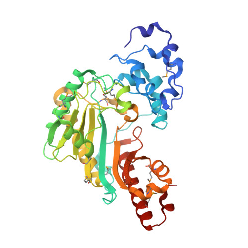
    Phosphagen kinase (PK) family members catalyze the reversible phosphoryl transfer between phosphagen and ADP to reserve or release energy in cell energy metabolism. The structures of classic quaternary complexes of dimeric creatine kinase (CK) revealed asymmetric ligand binding states of two protomers, but the significance and mechanism remain unclear. To understand this negative cooperativity further, we determined the first structure of dimeric arginine kinase (dAK), another PK family member, at 1.75 A, as well as the structure of its ternary complex with AMPPNP and arginine. Further structural analysis shows that the ligand-free protomer in a ligand-bound dimer opens more widely than the protomers in a ligand-free dimer, which leads to three different states of a dAK protomer. The unexpected allostery of the ligand-free protomer in a ligand-bound dimer should be relayed from the ligand-binding-induced allostery of its adjacent protomer. Mutations that weaken the interprotomer connections dramatically reduced the catalytic activities of dAK, indicating the importance of the allosteric propagation mediated by the homodimer interface. These results suggest a reciprocating mechanism of dimeric PK, which is shared by other ATP related oligomeric enzymes, e.g., ATP synthase.


  • Organizational Affiliation

    Laboratory of Structural Biology, Tsinghua University, Beijing, China.


Macromolecules
Find similar proteins by:  (by identity cutoff)  |  3D Structure
Entity ID: 1
MoleculeChains Sequence LengthOrganismDetailsImage
Arginine kinase
A, B, C, D
370Apostichopus japonicusMutation(s): 0 
Gene Names: AK
EC: 2.7.3.3
UniProt
Find proteins for Q9XY07 (Stichopus japonicus)
Explore Q9XY07 
Go to UniProtKB:  Q9XY07
Entity Groups  
Sequence Clusters30% Identity50% Identity70% Identity90% Identity95% Identity100% Identity
UniProt GroupQ9XY07
Sequence Annotations
Expand
  • Reference Sequence
Small Molecules
Ligands 2 Unique
IDChains Name / Formula / InChI Key2D Diagram3D Interactions
ANP
Query on ANP

Download Ideal Coordinates CCD File 
F [auth A]PHOSPHOAMINOPHOSPHONIC ACID-ADENYLATE ESTER
C10 H17 N6 O12 P3
PVKSNHVPLWYQGJ-KQYNXXCUSA-N
ARG
Query on ARG

Download Ideal Coordinates CCD File 
E [auth A]ARGININE
C6 H15 N4 O2
ODKSFYDXXFIFQN-BYPYZUCNSA-O
Modified Residues  1 Unique
IDChains TypeFormula2D DiagramParent
MSE
Query on MSE
A, B, C, D
L-PEPTIDE LINKINGC5 H11 N O2 SeMET
Experimental Data & Validation

Experimental Data

  • Method: X-RAY DIFFRACTION
  • Resolution: 2.45 Å
  • R-Value Free: 0.254 
  • R-Value Work: 0.214 
  • R-Value Observed: 0.223 
  • Space Group: P 1 21 1
Unit Cell:
Length ( Å )Angle ( ˚ )
a = 86.823α = 90
b = 60.073β = 93.08
c = 139.946γ = 90
Software Package:
Software NamePurpose
MAR345dtbdata collection
CNSrefinement
HKL-2000data reduction
HKL-2000data scaling
CNSphasing

Structure Validation

View Full Validation Report


Ligand Structure Quality Assessment 


Entry History 

Deposition Data

Revision History  (Full details and data files)

  • Version 1.0: 2009-09-29
    Type: Initial release
  • Version 1.1: 2011-07-13
    Changes: Version format compliance
  • Version 1.2: 2023-11-01
    Changes: Data collection, Database references, Derived calculations, Refinement description
  • Version 1.3: 2023-11-22
    Changes: Data collection