3JT1

Legionella pneumophila glucosyltransferase Lgt1, UDP-bound form


Experimental Data Snapshot

  • Method: X-RAY DIFFRACTION
  • Resolution: 2.30 Å
  • R-Value Free: 0.262 
  • R-Value Work: 0.185 
  • R-Value Observed: 0.189 

wwPDB Validation   3D Report Full Report


Ligand Structure Quality Assessment 


This is version 1.2 of the entry. See complete history


Literature

Structural Basis of the Action of Glucosyltransferase Lgt1 from Legionella pneumophila.

Lu, W.Du, J.Stahl, M.Tzivelekidis, T.Belyi, Y.Gerhardt, S.Aktories, K.Einsle, O.

(2009) J Mol Biol 

  • DOI: https://doi.org/10.1016/j.jmb.2009.11.044
  • Primary Citation of Related Structures:  
    3JSZ, 3JT1

  • PubMed Abstract: 

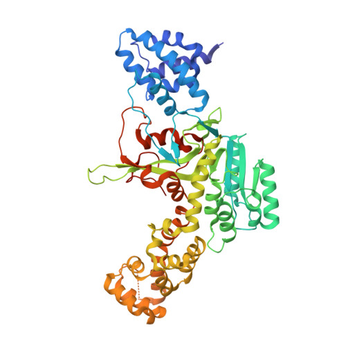
    The glucosyltransferase Lgt1 is one of three glucosylating toxins of Legionella pneumophila, the causative agent of Legionnaires disease. It acts through specific glucosylation of a serine residue (S53) in the eukaryotic elongation factor 1A and belongs to type A glycosyltransferases. High-resolution crystal structures of Lgt1 show an elongated shape of the protein, with the binding site for uridine disphosphate glucose at the bottom of a deep cleft. Lgt1 shows only a low sequence identity with other type A glycosyltransferases, and structural conservation is limited to a central folding core that is usually observed within this family of proteins. Domains and protrusions added to the core motif represent determinants for the specific recognition and binding of the target. Manual docking experiments based on the crystal structures of toxin and target protein suggest an obvious mode of binding to the target that allows for efficient transfer of a glucose moiety.


  • Organizational Affiliation

    Institut für organische Chemie und Biochemie, Albert-Ludwigs-Universität Freiburg, D-79104 Freiburg, Germany.


Macromolecules
Find similar proteins by:  (by identity cutoff)  |  3D Structure
Entity ID: 1
MoleculeChains Sequence LengthOrganismDetailsImage
Putative uncharacterized protein525Legionella pneumophila str. LensMutation(s): 0 
UniProt
Find proteins for Q5WWY0 (Legionella pneumophila (strain Lens))
Explore Q5WWY0 
Go to UniProtKB:  Q5WWY0
Entity Groups  
Sequence Clusters30% Identity50% Identity70% Identity90% Identity95% Identity100% Identity
UniProt GroupQ5WWY0
Sequence Annotations
Expand
  • Reference Sequence
Small Molecules
Ligands 1 Unique
IDChains Name / Formula / InChI Key2D Diagram3D Interactions
UDP
Query on UDP

Download Ideal Coordinates CCD File 
B [auth A]URIDINE-5'-DIPHOSPHATE
C9 H14 N2 O12 P2
XCCTYIAWTASOJW-XVFCMESISA-N
Experimental Data & Validation

Experimental Data

  • Method: X-RAY DIFFRACTION
  • Resolution: 2.30 Å
  • R-Value Free: 0.262 
  • R-Value Work: 0.185 
  • R-Value Observed: 0.189 
  • Space Group: H 3
Unit Cell:
Length ( Å )Angle ( ˚ )
a = 122.571α = 90
b = 122.571β = 90
c = 103.982γ = 120
Software Package:
Software NamePurpose
REFMACrefinement

Structure Validation

View Full Validation Report


Ligand Structure Quality Assessment 


Entry History 

Deposition Data

Revision History  (Full details and data files)

  • Version 1.0: 2010-02-02
    Type: Initial release
  • Version 1.1: 2011-07-13
    Changes: Advisory, Version format compliance
  • Version 1.2: 2023-11-01
    Changes: Data collection, Database references, Derived calculations, Refinement description