3JC2

The structure of the mammalian Sec61 channel opened by a signal sequence


Experimental Data Snapshot

  • Method: ELECTRON MICROSCOPY
  • Resolution: 3.60 Å
  • Aggregation State: PARTICLE 
  • Reconstruction Method: SINGLE PARTICLE 

wwPDB Validation   3D Report Full Report


This is version 1.1 of the entry. See complete history


Literature

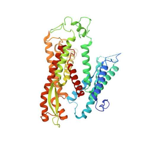
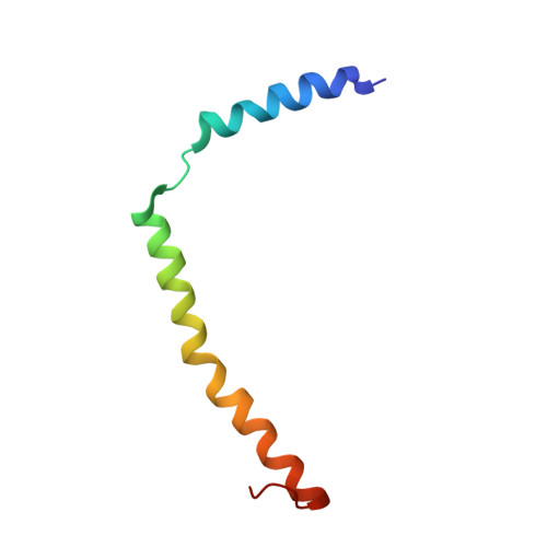
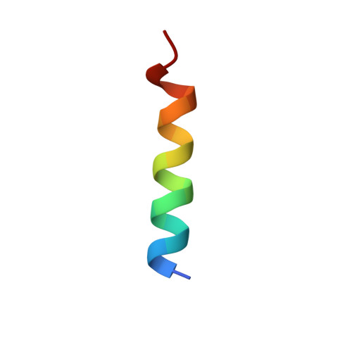
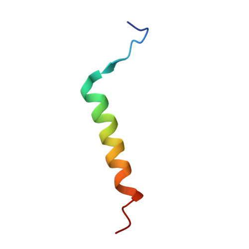
Structure of the Sec61 channel opened by a signal sequence.

Voorhees, R.M.Hegde, R.S.

(2016) Science 351: 88-91

  • DOI: https://doi.org/10.1126/science.aad4992
  • Primary Citation of Related Structures:  
    3JC2

  • PubMed Abstract: 

    Secreted and integral membrane proteins compose up to one-third of the biological proteome. These proteins contain hydrophobic signals that direct their translocation across or insertion into the lipid bilayer by the Sec61 protein-conducting channel. The molecular basis of how hydrophobic signals within a nascent polypeptide trigger channel opening is not understood. Here, we used cryo-electron microscopy to determine the structure of an active Sec61 channel that has been opened by a signal sequence. The signal supplants helix 2 of Sec61α, which triggers a rotation that opens the central pore both axially across the membrane and laterally toward the lipid bilayer. Comparisons with structures of Sec61 in other states suggest a pathway for how hydrophobic signals engage the channel to gain access to the lipid bilayer.


  • Organizational Affiliation

    MRC Laboratory of Molecular Biology, Medical Research Council, Francis Crick Avenue, Cambridge CB2 0QH, UK.


Macromolecules
Find similar proteins by:  (by identity cutoff)  |  3D Structure
Entity ID: 1
MoleculeChains Sequence LengthOrganismDetailsImage
Protein transport protein Sec61 subunit alpha isoform 1A [auth 1]476Canis lupus familiarisMutation(s): 0 
Membrane Entity: Yes 
UniProt
Find proteins for P38377 (Canis lupus familiaris)
Explore P38377 
Go to UniProtKB:  P38377
Entity Groups  
Sequence Clusters30% Identity50% Identity70% Identity90% Identity95% Identity100% Identity
UniProt GroupP38377
Sequence Annotations
Expand
  • Reference Sequence
Find similar proteins by:  (by identity cutoff)  |  3D Structure
Entity ID: 2
MoleculeChains Sequence LengthOrganismDetailsImage
Protein transport protein Sec61 subunit gammaB [auth 2]62Canis lupus familiarisMutation(s): 0 
Membrane Entity: Yes 
UniProt
Find proteins for P60058 (Canis lupus familiaris)
Explore P60058 
Go to UniProtKB:  P60058
Entity Groups  
Sequence Clusters30% Identity50% Identity70% Identity90% Identity95% Identity100% Identity
UniProt GroupP60058
Sequence Annotations
Expand
  • Reference Sequence

Find similar proteins by:  Sequence   |   3D Structure  

Entity ID: 3
MoleculeChains Sequence LengthOrganismDetailsImage
ProlactinC [auth w]19Bos taurusMutation(s): 0 
Gene Names: prolactin
Membrane Entity: Yes 
UniProt
Find proteins for P01239 (Bos taurus)
Explore P01239 
Go to UniProtKB:  P01239
Entity Groups  
Sequence Clusters30% Identity50% Identity70% Identity90% Identity95% Identity100% Identity
UniProt GroupP01239
Sequence Annotations
Expand
  • Reference Sequence
Find similar proteins by:  (by identity cutoff)  |  3D Structure
Entity ID: 4
MoleculeChains Sequence LengthOrganismDetailsImage
Protein transport protein Sec61 subunit betaD [auth 3]32Canis lupus familiarisMutation(s): 0 
Sequence Annotations
Expand
  • Reference Sequence
Experimental Data & Validation

Experimental Data

  • Method: ELECTRON MICROSCOPY
  • Resolution: 3.60 Å
  • Aggregation State: PARTICLE 
  • Reconstruction Method: SINGLE PARTICLE 
EM Software:
TaskSoftware PackageVersion
RECONSTRUCTIONRELION

Structure Validation

View Full Validation Report


Entry History 

Deposition Data

Revision History  (Full details and data files)

  • Version 1.0: 2016-01-13
    Type: Initial release
  • Version 1.1: 2018-07-18
    Changes: Data collection