3IWT

Structure of hypothetical molybdenum cofactor biosynthesis protein B from Sulfolobus tokodaii


Experimental Data Snapshot

  • Method: X-RAY DIFFRACTION
  • Resolution: 1.90 Å
  • R-Value Free: 0.187 
  • R-Value Work: 0.168 
  • R-Value Observed: 0.169 

wwPDB Validation   3D Report Full Report


This is version 1.3 of the entry. See complete history


Literature

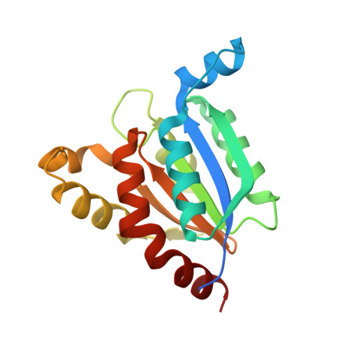
Structure of hypothetical Mo-cofactor biosynthesis protein B (ST2315) from Sulfolobus tokodaii

Antonyuk, S.V.Strange, R.W.Ellis, M.J.Bessho, Y.Kuramitsu, S.Shinkai, A.Yokoyama, S.Hasnain, S.S.

(2009) Acta Crystallogr Sect F Struct Biol Cryst Commun 65: 1200-1203

  • DOI: https://doi.org/10.1107/S1744309109043772
  • Primary Citation of Related Structures:  
    3IWT

  • PubMed Abstract: 

    The structure of a probable Mo-cofactor biosynthesis protein B from Sulfolobus tokodaii, belonging to space group P6(4)22 with unit-cell parameters a = b = 136.68, c = 210.52 A, was solved by molecular replacement to a resolution of 1.9 A and refined to an R factor and R(free) of 16.8% and 18.5%, respectively. The asymmetric unit contains a trimer, while the biologically significant oligomer is predicted to be a hexamer by size-exclusion chromatography. The subunit structure and fold of ST2315 are similar to those of other enzymes that are known to be involved in the molybdopterin- and molybdenum cofactor-biosynthesis pathways.


  • Organizational Affiliation

    Molecular Biophysics Group, School of Biological Sciences, University of Liverpool, Crown Street, Liverpool L69 7ZB, England.


Macromolecules
Find similar proteins by:  (by identity cutoff)  |  3D Structure
Entity ID: 1
MoleculeChains Sequence LengthOrganismDetailsImage
178aa long hypothetical molybdenum cofactor biosynthesis protein B
A, B, C
178Sulfurisphaera tokodaiiMutation(s): 0 
Gene Names: ST2315
UniProt
Find proteins for Q96Y52 (Sulfurisphaera tokodaii (strain DSM 16993 / JCM 10545 / NBRC 100140 / 7))
Explore Q96Y52 
Go to UniProtKB:  Q96Y52
Entity Groups  
Sequence Clusters30% Identity50% Identity70% Identity90% Identity95% Identity100% Identity
UniProt GroupQ96Y52
Sequence Annotations
Expand
  • Reference Sequence
Small Molecules
Ligands 3 Unique
IDChains Name / Formula / InChI Key2D Diagram3D Interactions
PEG
Query on PEG

Download Ideal Coordinates CCD File 
D [auth A]
H [auth B]
I [auth B]
K [auth B]
L [auth B]
D [auth A],
H [auth B],
I [auth B],
K [auth B],
L [auth B],
M [auth B],
N [auth B],
O [auth C]
DI(HYDROXYETHYL)ETHER
C4 H10 O3
MTHSVFCYNBDYFN-UHFFFAOYSA-N
GOL
Query on GOL

Download Ideal Coordinates CCD File 
F [auth A],
J [auth B]
GLYCEROL
C3 H8 O3
PEDCQBHIVMGVHV-UHFFFAOYSA-N
MG
Query on MG

Download Ideal Coordinates CCD File 
E [auth A],
G [auth B]
MAGNESIUM ION
Mg
JLVVSXFLKOJNIY-UHFFFAOYSA-N
Experimental Data & Validation

Experimental Data

  • Method: X-RAY DIFFRACTION
  • Resolution: 1.90 Å
  • R-Value Free: 0.187 
  • R-Value Work: 0.168 
  • R-Value Observed: 0.169 
  • Space Group: P 64 2 2
Unit Cell:
Length ( Å )Angle ( ˚ )
a = 136.675α = 90
b = 136.675β = 90
c = 210.525γ = 120
Software Package:
Software NamePurpose
DENZOdata reduction
SCALEPACKdata scaling
MOLREPphasing
REFMACrefinement
PDB_EXTRACTdata extraction
HKL-2000data collection

Structure Validation

View Full Validation Report


Entry History 

Deposition Data

Revision History  (Full details and data files)

  • Version 1.0: 2009-09-29
    Type: Initial release
  • Version 1.1: 2011-07-13
    Changes: Advisory, Version format compliance
  • Version 1.2: 2017-11-01
    Changes: Refinement description
  • Version 1.3: 2023-11-01
    Changes: Data collection, Database references, Derived calculations, Refinement description