3ISN

Crystal structure of HIV-1 RT bound to A 6-vinylpyrimidine inhibitor


Experimental Data Snapshot

  • Method: X-RAY DIFFRACTION
  • Resolution: 2.50 Å
  • R-Value Free: 0.281 
  • R-Value Work: 0.221 
  • R-Value Observed: 0.224 

wwPDB Validation   3D Report Full Report


Ligand Structure Quality Assessment 


This is version 1.2 of the entry. See complete history


Literature

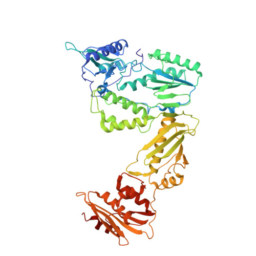
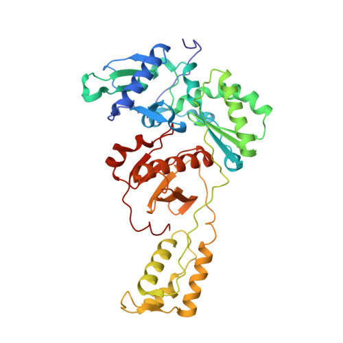
Crystal Structure of HIV-1 Reverse Transcriptase Bound to a Non-Nucleoside Inhibitor with a Novel Mechanism of Action

Freisz, S.Bec, G.Radi, M.Wolff, P.Crespan, E.Angeli, L.Dumas, P.Maga, G.Botta, M.Ennifar, E.

(2010) Angew Chem Int Ed Engl 49: 1805-1808


Macromolecules
Find similar proteins by:  (by identity cutoff)  |  3D Structure
Entity ID: 1
MoleculeChains Sequence LengthOrganismDetailsImage
Reverse transcriptase/ribonuclease HA [auth C]560Human immunodeficiency virus type 1 BH10Mutation(s): 0 
Gene Names: gag-pol
EC: 2.7.7.49 (PDB Primary Data), 2.7.7.7 (PDB Primary Data), 3.1.26.4 (PDB Primary Data)
UniProt
Find proteins for P03366 (Human immunodeficiency virus type 1 group M subtype B (isolate BH10))
Explore P03366 
Go to UniProtKB:  P03366
Entity Groups  
Sequence Clusters30% Identity50% Identity70% Identity90% Identity95% Identity100% Identity
UniProt GroupP03366
Sequence Annotations
Expand
  • Reference Sequence
Find similar proteins by:  (by identity cutoff)  |  3D Structure
Entity ID: 2
MoleculeChains Sequence LengthOrganismDetailsImage
p51 RTB [auth D]427Human immunodeficiency virus type 1 BH10Mutation(s): 0 
UniProt
Find proteins for P03366 (Human immunodeficiency virus type 1 group M subtype B (isolate BH10))
Explore P03366 
Go to UniProtKB:  P03366
Entity Groups  
Sequence Clusters30% Identity50% Identity70% Identity90% Identity95% Identity100% Identity
UniProt GroupP03366
Sequence Annotations
Expand
  • Reference Sequence
Small Molecules
Ligands 1 Unique
IDChains Name / Formula / InChI Key2D Diagram3D Interactions
EDM
Query on EDM

Download Ideal Coordinates CCD File 
C
6-ethenyl-N,N-dimethyl-2-(methylsulfonyl)pyrimidin-4-amine
C9 H13 N3 O2 S
LWXYVFFCCMAZFC-UHFFFAOYSA-N
Binding Affinity Annotations 
IDSourceBinding Affinity
EDM Binding MOAD:  3ISN Ki: 8 (nM) from 1 assay(s)
Experimental Data & Validation

Experimental Data

  • Method: X-RAY DIFFRACTION
  • Resolution: 2.50 Å
  • R-Value Free: 0.281 
  • R-Value Work: 0.221 
  • R-Value Observed: 0.224 
  • Space Group: C 1 2 1
Unit Cell:
Length ( Å )Angle ( ˚ )
a = 225.121α = 90
b = 72.182β = 108.97
c = 93.516γ = 90
Software Package:
Software NamePurpose
MOLREPphasing
PHENIXrefinement
XDSdata reduction
XSCALEdata scaling

Structure Validation

View Full Validation Report


Ligand Structure Quality Assessment 


Entry History 

Deposition Data

Revision History  (Full details and data files)

  • Version 1.0: 2010-03-16
    Type: Initial release
  • Version 1.1: 2011-07-13
    Changes: Version format compliance
  • Version 1.2: 2023-11-01
    Changes: Data collection, Database references, Derived calculations, Refinement description