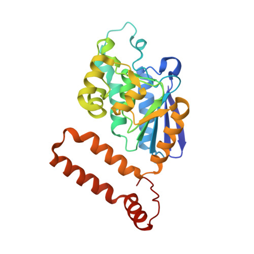
Crystal structure of phenylacetic acid degradation protein PaaG from Thermus thermophilus HB8
Kichise, T., Hisano, T., Takeda, K., Miki, K.(2009) Proteins 76: 779-786
- PubMed: 19452559
- DOI: https://doi.org/10.1002/prot.22455
- Primary Citation of Related Structures:
3HRX - PubMed Abstract:
Microbial degradation of phenylacetic acid proceeds via the hybrid pathway that includes formation of a coenzyme A thioester, ring hydroxylation, non-oxygenolytic ring opening, and beta-oxidation-like reactions. A phenylacetic acid degradation protein PaaG is a member of the crotonase superfamily, and is a candidate non-oxygenolytic ring-opening enzyme. The crystal structure of PaaG from Thermus thermophilus HB8 was determined at a resolution of 1.85 A. PaaG consists of three identical subunits related by local three-fold symmetry. The monomer is comprised of a spiral and a helical domain with a fold characteristic of the crotonase superfamily. A putative active site residue, Asp136, is situated in an active site cavity and surrounded by several hydrophobic and hydrophilic residues. The active site cavity is sufficiently large to accommodate a ring substrate. Two conformations are observed for helix H2 located adjacent to the active site. Helix H2 is kinked at Asn81 in two subunits, whereas it is kinked at Leu77 in the other subunit, and the side chain of Tyr80 is closer to Asp136. This indicates that catalytic reaction of PaaG may proceed with large conformational changes at the active site. Asp136 is the only conserved polar residue in the active site. It is located at the same position as those of 4-chlorobenzoyl-CoA dehalogenase and peroxisomal Delta(3),Delta(2)-enoyl-CoA isomerase, indicating that PaaG may undergo isomerization or a ring-opening reaction via a Delta(3),Delta(2)-enoyl-CoA isomerase-like mechanism.
Organizational Affiliation:
RIKEN SPring-8 Center at Harima Institute, Koto 1-1-1, Sayo-cho, Sayo-gun, Hyogo 679-5148, Japan.