3H5B

Crystal structure of wild type HIV-1 protease with novel P1'-ligand GRL-02031


Experimental Data Snapshot

  • Method: X-RAY DIFFRACTION
  • Resolution: 1.29 Å
  • R-Value Free: 0.181 
  • R-Value Work: 0.142 
  • R-Value Observed: 0.144 

wwPDB Validation   3D Report Full Report


Ligand Structure Quality Assessment 


This is version 1.4 of the entry. See complete history


Literature

Design of HIV-1 protease inhibitors with pyrrolidinones and oxazolidinones as novel P1'-ligands to enhance backbone-binding interactions with protease: synthesis, biological evaluation, and protein-ligand X-ray studies.

Ghosh, A.K.Leshchenko-Yashchuk, S.Anderson, D.D.Baldridge, A.Noetzel, M.Miller, H.B.Tie, Y.Wang, Y.F.Koh, Y.Weber, I.T.Mitsuya, H.

(2009) J Med Chem 52: 3902-3914

  • DOI: https://doi.org/10.1021/jm900303m
  • Primary Citation of Related Structures:  
    3H5B

  • PubMed Abstract: 

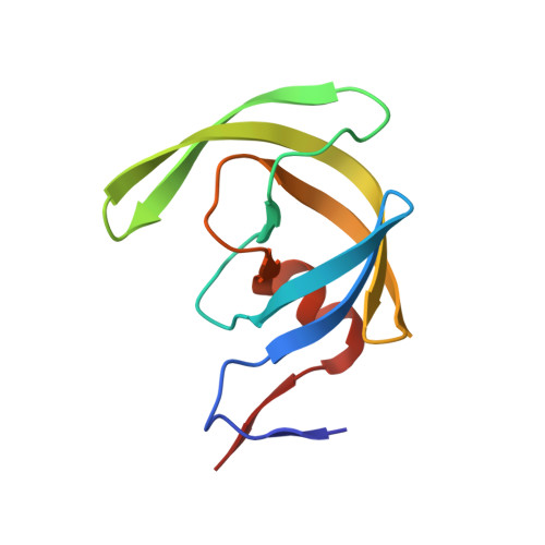
    Structure-based design, synthesis, and biological evaluation of a series of novel HIV-1 protease inhibitors are described. In an effort to enhance interactions with protease backbone atoms, we have incorporated stereochemically defined methyl-2-pyrrolidinone and methyl oxazolidinone as the P1'-ligands. These ligands are designed to interact with Gly-27' carbonyl and Arg-8 side chain in the S1'-subsite of the HIV protease. We have investigated the potential of these ligands in combination with our previously developed bis-tetrahydrofuran (bis-THF) and cyclopentanyltetrahydrofuran (Cp-THF) as the P2-ligands. Inhibitor 19b with a (R)-aminomethyl-2-pyrrolidinone and a Cp-THF was shown to be the most potent compound. This inhibitor maintained near full potency against multi-PI-resistant clinical HIV-1 variants. A high resolution protein-ligand X-ray crystal structure of 19b-bound HIV-1 protease revealed that the P1'-pyrrolidinone heterocycle and the P2-Cp-ligand are involved in several critical interactions with the backbone atoms in the S1' and S2 subsites of HIV-1 protease.


  • Organizational Affiliation

    Department of Chemistry, Purdue University, West Lafayette, Indiana 47907, USA. akghosh@purdue.edu


Macromolecules
Find similar proteins by:  (by identity cutoff)  |  3D Structure
Entity ID: 1
MoleculeChains Sequence LengthOrganismDetailsImage
HIV-1 protease
A, B
99Human immunodeficiency virus type 1 (BRU ISOLATE)Mutation(s): 5 
Gene Names: gag-pol
EC: 3.4.23.16
UniProt
Find proteins for P03367 (Human immunodeficiency virus type 1 group M subtype B (isolate BRU/LAI))
Explore P03367 
Go to UniProtKB:  P03367
Entity Groups  
Sequence Clusters30% Identity50% Identity70% Identity90% Identity95% Identity100% Identity
UniProt GroupP03367
Sequence Annotations
Expand
  • Reference Sequence
Small Molecules
Binding Affinity Annotations 
IDSourceBinding Affinity
031 BindingDB:  3H5B Ki: min: 0.1, max: 0.44 (nM) from 2 assay(s)
Binding MOAD:  3H5B Ki: 0.1 (nM) from 1 assay(s)
Experimental Data & Validation

Experimental Data

  • Method: X-RAY DIFFRACTION
  • Resolution: 1.29 Å
  • R-Value Free: 0.181 
  • R-Value Work: 0.142 
  • R-Value Observed: 0.144 
  • Space Group: P 21 21 2
Unit Cell:
Length ( Å )Angle ( ˚ )
a = 58.111α = 90
b = 86.416β = 90
c = 45.97γ = 90
Software Package:
Software NamePurpose
MAR345data collection
AMoREphasing
SHELXL-97refinement
HKL-2000data reduction
HKL-2000data scaling

Structure Validation

View Full Validation Report


Ligand Structure Quality Assessment 


Entry History 

Deposition Data

Revision History  (Full details and data files)

  • Version 1.0: 2009-06-16
    Type: Initial release
  • Version 1.1: 2011-07-13
    Changes: Non-polymer description, Version format compliance
  • Version 1.2: 2017-11-01
    Changes: Refinement description
  • Version 1.3: 2021-10-13
    Changes: Database references, Derived calculations
  • Version 1.4: 2023-09-06
    Changes: Data collection, Refinement description