3FP5

Crystal structure of ACBP from Moniliophthora perniciosa


Experimental Data Snapshot

  • Method: X-RAY DIFFRACTION
  • Resolution: 1.61 Å
  • R-Value Free: 0.212 
  • R-Value Work: 0.186 
  • R-Value Observed: 0.188 

wwPDB Validation   3D Report Full Report


This is version 1.2 of the entry. See complete history


Literature

A new topology of ACBP from Moniliophthora perniciosa.

Monzani, P.S.Pereira, H.M.Melo, F.A.Meirelles, F.V.Oliva, G.Cascardo, J.C.

(2010) Biochim Biophys Acta 1804: 115-123

  • DOI: https://doi.org/10.1016/j.bbapap.2009.09.020
  • Primary Citation of Related Structures:  
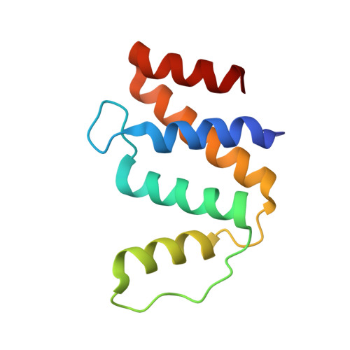
    3FP5

  • PubMed Abstract: 

    Acyl-CoA binding protein (ACBP) is a housekeeping protein and is an essential protein in human cell lines and in Trypanosoma brucei. The ACBP of Moniliophthora perniciosa is composed of 104 amino acids and is possibly a non-classic isoform exclusively from Basidiomycetes. The M. perniciosa acbp gene was cloned, and the protein was expressed and purified. Acyl-CoA ester binding was analyzed by isoelectric focusing, native gel electrophoresis and isothermal titration calorimetry. Our results suggest an increasing affinity of ACBP for longer acyl-CoA esters, such as myristoyl-CoA to arachidoyl-CoA, and best fit modeling indicates two binding sites. ACBP undergoes a shift from a monomeric to a dimeric state, as shown by dynamic light scattering, fluorescence anisotropy and native gel electrophoresis in the absence and presence of the ligand. The protein's structure was determined at 1.6 A resolution and revealed a new topology for ACBP, containing five alpha-helices instead of four. alpha-helices 1, 2, 3 and 4 adopted a bundled arrangement that is unique from the previously determined four-helix folds of ACBP, while alpha-helices 1, 2, 4 and 5 formed a classical four-helix bundle. A MES molecule was found in the CoA binding site, suggesting that the CoA site could be a target for small compound screening.


  • Organizational Affiliation

    Departamento de Ciências Biológicas, Laboratório de Proteômica, Centro de Biotecnologia e Genética, Universidade Estadual de Santa Cruz, Ilhéus, BA, CEP 45662-900, Brazil.


Macromolecules
Find similar proteins by:  (by identity cutoff)  |  3D Structure
Entity ID: 1
MoleculeChains Sequence LengthOrganismDetailsImage
Acyl-CoA Binding Protein106Moniliophthora perniciosaMutation(s): 0 
Gene Names: MpACBP
UniProt
Find proteins for D1MPT1 (Moniliophthora perniciosa)
Explore D1MPT1 
Go to UniProtKB:  D1MPT1
Entity Groups  
Sequence Clusters30% Identity50% Identity70% Identity90% Identity95% Identity100% Identity
UniProt GroupD1MPT1
Sequence Annotations
Expand
  • Reference Sequence
Small Molecules
Ligands 2 Unique
IDChains Name / Formula / InChI Key2D Diagram3D Interactions
MES
Query on MES

Download Ideal Coordinates CCD File 
C [auth A]2-(N-MORPHOLINO)-ETHANESULFONIC ACID
C6 H13 N O4 S
SXGZJKUKBWWHRA-UHFFFAOYSA-N
ZN
Query on ZN

Download Ideal Coordinates CCD File 
B [auth A]ZINC ION
Zn
PTFCDOFLOPIGGS-UHFFFAOYSA-N
Experimental Data & Validation

Experimental Data

  • Method: X-RAY DIFFRACTION
  • Resolution: 1.61 Å
  • R-Value Free: 0.212 
  • R-Value Work: 0.186 
  • R-Value Observed: 0.188 
  • Space Group: P 21 21 21
Unit Cell:
Length ( Å )Angle ( ˚ )
a = 32.5α = 90
b = 41.024β = 90
c = 82.463γ = 90
Software Package:
Software NamePurpose
SCALAdata scaling
PHASERphasing
PHENIXrefinement
PDB_EXTRACTdata extraction
MOSFLMdata reduction

Structure Validation

View Full Validation Report


Entry History 

Deposition Data

Revision History  (Full details and data files)

  • Version 1.0: 2009-11-17
    Type: Initial release
  • Version 1.1: 2011-07-13
    Changes: Version format compliance
  • Version 1.2: 2023-09-06
    Changes: Data collection, Database references, Derived calculations, Refinement description