3FOW

Plasmodium Purine Nucleoside Phosphorylase V66I-V73I-Y160F Mutant


Experimental Data Snapshot

  • Method: X-RAY DIFFRACTION
  • Resolution: 2.80 Å
  • R-Value Free: 0.202 
  • R-Value Work: 0.165 
  • R-Value Observed: 0.167 

wwPDB Validation   3D Report Full Report


Ligand Structure Quality Assessment 


This is version 2.1 of the entry. See complete history


Literature

Structural determinants of the 5'-methylthioinosine specificity of Plasmodium purine nucleoside phosphorylase.

Donaldson, T.M.Ting, L.M.Zhan, C.Shi, W.Zheng, R.Almo, S.C.Kim, K.

(2014) PLoS One 9: e84384-e84384

  • DOI: https://doi.org/10.1371/journal.pone.0084384
  • Primary Citation of Related Structures:  
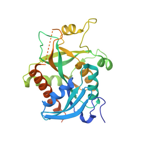
    3FOW

  • PubMed Abstract: 

    Plasmodium parasites rely upon purine salvage for survival. Plasmodium purine nucleoside phosphorylase is part of the streamlined Plasmodium purine salvage pathway that leads to the phosphorylysis of both purines and 5'-methylthiopurines, byproducts of polyamine synthesis. We have explored structural features in Plasmodium falciparum purine nucleoside phosphorylase (PfPNP) that affect efficiency of catalysis as well as those that make it suitable for dual specificity. We used site directed mutagenesis to identify residues critical for PfPNP catalytic activity as well as critical residues within a hydrophobic pocket required for accommodation of the 5'-methylthio group. Kinetic analysis data shows that several mutants had disrupted binding of the 5'-methylthio group while retaining activity for inosine. A triple PfPNP mutant that mimics Toxoplasma gondii PNP had significant loss of 5'-methylthio activity with retention of inosine activity. Crystallographic investigation of the triple mutant PfPNP with Tyr160Phe, Val66Ile, andVal73Ile in complex with the transition state inhibitor immucillin H reveals fewer hydrogen bond interactions for the inhibitor in the hydrophobic pocket.


  • Organizational Affiliation

    Departments of Medicine, Pathology, and of Microbiology & Immunology, Albert Einstein College of Medicine, Bronx, New York, United States of America.


Macromolecules
Find similar proteins by:  (by identity cutoff)  |  3D Structure
Entity ID: 1
MoleculeChains Sequence LengthOrganismDetailsImage
Uridine phosphorylase, putative
A, B
276Plasmodium falciparum 3D7Mutation(s): 3 
Gene Names: PFE0660cPNP
EC: 2.4.2.3
UniProt
Find proteins for Q8I3X4 (Plasmodium falciparum (isolate 3D7))
Explore Q8I3X4 
Go to UniProtKB:  Q8I3X4
Entity Groups  
Sequence Clusters30% Identity50% Identity70% Identity90% Identity95% Identity100% Identity
UniProt GroupQ8I3X4
Sequence Annotations
Expand
  • Reference Sequence
Experimental Data & Validation

Experimental Data

  • Method: X-RAY DIFFRACTION
  • Resolution: 2.80 Å
  • R-Value Free: 0.202 
  • R-Value Work: 0.165 
  • R-Value Observed: 0.167 
  • Space Group: I 41 3 2
Unit Cell:
Length ( Å )Angle ( ˚ )
a = 234.969α = 90
b = 234.969β = 90
c = 234.969γ = 90
Software Package:
Software NamePurpose
CBASSdata collection
PHASERphasing
REFMACrefinement
HKL-2000data reduction
HKL-2000data scaling

Structure Validation

View Full Validation Report


Ligand Structure Quality Assessment 


Entry History 

Deposition Data

Revision History  (Full details and data files)

  • Version 1.0: 2009-12-29
    Type: Initial release
  • Version 1.1: 2011-07-13
    Changes: Source and taxonomy, Version format compliance
  • Version 1.2: 2014-03-05
    Changes: Database references
  • Version 1.3: 2021-10-20
    Changes: Database references, Derived calculations
  • Version 2.0: 2022-10-05
    Changes: Atomic model, Structure summary
  • Version 2.1: 2023-09-20
    Changes: Data collection, Refinement description