3FLF

Thermolysin inhibition


Experimental Data Snapshot

  • Method: X-RAY DIFFRACTION
  • Resolution: 1.97 Å
  • R-Value Free: 0.265 
  • R-Value Work: 0.200 
  • R-Value Observed: 0.202 

wwPDB Validation   3D Report Full Report


Ligand Structure Quality Assessment 


This is version 1.6 of the entry. See complete history


Literature

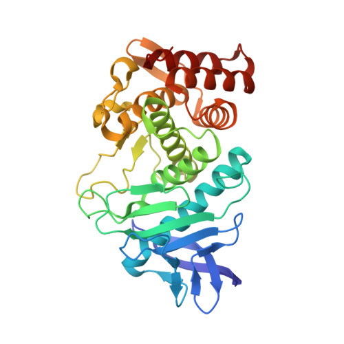
Displacement of disordered water molecules from hydrophobic pocket creates enthalpic signature: binding of phosphonamidate to the S1'-pocket of thermolysin.

Englert, L.Biela, A.Zayed, M.Heine, A.Hangauer, D.Klebe, G.

(2010) Biochim Biophys Acta 1800: 1192-1202

  • DOI: https://doi.org/10.1016/j.bbagen.2010.06.009
  • Primary Citation of Related Structures:  
    3FLF, 3FV4

  • PubMed Abstract: 

    Prerequisite for the design of tight binding protein inhibitors and prediction of their properties is an in-depth understanding of the structural and thermodynamic details of the binding process. A series of closely related phosphonamidates was studied to elucidate the forces underlying their binding affinity to thermolysin. The investigated inhibitors are identical except for the parts penetrating into the hydrophobic S₁'-pocket.


  • Organizational Affiliation

    Department of Pharmaceutical Chemistry, Philipps-University Marburg, 35032 Marburg, Germany.


Macromolecules
Find similar proteins by:  (by identity cutoff)  |  3D Structure
Entity ID: 1
MoleculeChains Sequence LengthOrganismDetailsImage
Thermolysin316Bacillus thermoproteolyticusMutation(s): 0 
EC: 3.4.24.27
UniProt
Find proteins for P00800 (Bacillus thermoproteolyticus)
Explore P00800 
Go to UniProtKB:  P00800
Entity Groups  
Sequence Clusters30% Identity50% Identity70% Identity90% Identity95% Identity100% Identity
UniProt GroupP00800
Sequence Annotations
Expand
  • Reference Sequence
Small Molecules
Ligands 4 Unique
IDChains Name / Formula / InChI Key2D Diagram3D Interactions
UB3
Query on UB3

Download Ideal Coordinates CCD File 
G [auth A]N-[(S)-({[(benzyloxy)carbonyl]amino}methyl)(hydroxy)phosphoryl]-L-valyl-L-leucine
C20 H32 N3 O7 P
NFEZDCCCWWWKAJ-IRXDYDNUSA-N
GOL
Query on GOL

Download Ideal Coordinates CCD File 
H [auth A],
I [auth A]
GLYCEROL
C3 H8 O3
PEDCQBHIVMGVHV-UHFFFAOYSA-N
ZN
Query on ZN

Download Ideal Coordinates CCD File 
F [auth A]ZINC ION
Zn
PTFCDOFLOPIGGS-UHFFFAOYSA-N
CA
Query on CA

Download Ideal Coordinates CCD File 
B [auth A],
C [auth A],
D [auth A],
E [auth A]
CALCIUM ION
Ca
BHPQYMZQTOCNFJ-UHFFFAOYSA-N
Experimental Data & Validation

Experimental Data

  • Method: X-RAY DIFFRACTION
  • Resolution: 1.97 Å
  • R-Value Free: 0.265 
  • R-Value Work: 0.200 
  • R-Value Observed: 0.202 
  • Space Group: P 61 2 2
Unit Cell:
Length ( Å )Angle ( ˚ )
a = 92.658α = 90
b = 92.658β = 90
c = 130.477γ = 120
Software Package:
Software NamePurpose
SHELXL-97refinement
CNSrefinement
CrystalCleardata collection
HKL-2000data reduction
HKL-2000data scaling
CNSphasing

Structure Validation

View Full Validation Report


Ligand Structure Quality Assessment 


Entry History 

Deposition Data

Revision History  (Full details and data files)

  • Version 1.0: 2010-01-12
    Type: Initial release
  • Version 1.1: 2011-07-13
    Changes: Atomic model, Database references, Derived calculations, Non-polymer description, Structure summary, Version format compliance
  • Version 1.2: 2012-12-12
    Changes: Other
  • Version 1.3: 2014-01-15
    Changes: Database references
  • Version 1.4: 2018-01-24
    Changes: Structure summary
  • Version 1.5: 2019-07-24
    Changes: Data collection, Refinement description
  • Version 1.6: 2023-09-06
    Changes: Data collection, Database references, Derived calculations, Refinement description