3FD9

Crystal Structure of the transcriptional anti-activator ExsD from Pseudomonas aeruginosa


Experimental Data Snapshot

  • Method: X-RAY DIFFRACTION
  • Resolution: 2.60 Å
  • R-Value Free: 0.263 
  • R-Value Work: 0.213 
  • R-Value Observed: 0.215 

wwPDB Validation   3D Report Full Report


This is version 1.3 of the entry. See complete history


Literature

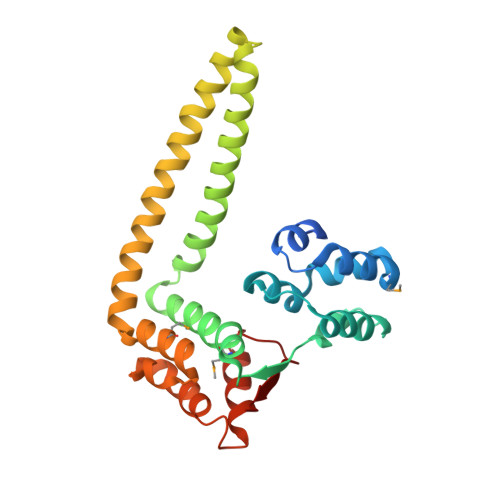
Structural evidence suggests that antiactivator ExsD from Pseudomonas aeruginosa is a DNA binding protein

Bernhards, R.C.Jing, X.Vogelaar, N.J.Robinson, H.Schubot, F.D.

(2009) Protein Sci 18: 503-513

  • DOI: https://doi.org/10.1002/pro.48
  • Primary Citation of Related Structures:  
    3FD9

  • PubMed Abstract: 

    The opportunistic pathogen P. aeruginosa utilizes a type III secretion system (T3SS) to support acute infections in predisposed individuals. In this bacterium, expression of all T3SS-related genes is dependent on the AraC-type transcriptional activator ExsA. Before host contact, the T3SS is inactive and ExsA is repressed by the antiactivator protein ExsD. The repression, thought to occur through direct interactions between the two proteins, is relieved upon opening of the type III secretion (T3S) channel when secretion chaperone ExsC sequesters ExsD. We have solved the crystal structure of Delta20ExsD, a protease-resistant fragment of ExsD that lacks only the 20 amino terminal residues of the wild-type protein at 2.6 A. Surprisingly the structure revealed similarities between ExsD and the DNA binding domain of transcriptional repressor KorB. A model of an ExsD-DNA complex constructed on the basis of this homology produced a realistic complex that is supported by the prevalence of conserved residues in the putative DNA binding site and the results of differential scanning fluorimetry studies. Our findings challenge the currently held model that ExsD solely acts through interactions with ExsA and raise new questions with respect to the underlying mechanism of ExsA regulation.


  • Organizational Affiliation

    Department of Biological Sciences, Virginia Polytechnic Institute and State University, Blacksburg, Virginia 24060, USA.


Macromolecules
Find similar proteins by:  (by identity cutoff)  |  3D Structure
Entity ID: 1
MoleculeChains Sequence LengthOrganismDetailsImage
Uncharacterized protein
A, B, C
256Pseudomonas aeruginosa UCBPP-PA14Mutation(s): 0 
Gene Names: exsDPA14_42380
UniProt
Find proteins for A0A0H2Z989 (Pseudomonas aeruginosa (strain UCBPP-PA14))
Explore A0A0H2Z989 
Go to UniProtKB:  A0A0H2Z989
Entity Groups  
Sequence Clusters30% Identity50% Identity70% Identity90% Identity95% Identity100% Identity
UniProt GroupA0A0H2Z989
Sequence Annotations
Expand
  • Reference Sequence
Small Molecules
Modified Residues  1 Unique
IDChains TypeFormula2D DiagramParent
MSE
Query on MSE
A, B, C
L-PEPTIDE LINKINGC5 H11 N O2 SeMET
Experimental Data & Validation

Experimental Data

  • Method: X-RAY DIFFRACTION
  • Resolution: 2.60 Å
  • R-Value Free: 0.263 
  • R-Value Work: 0.213 
  • R-Value Observed: 0.215 
  • Space Group: P 21 21 2
Unit Cell:
Length ( Å )Angle ( ˚ )
a = 144.755α = 90
b = 69.347β = 90
c = 86.331γ = 90
Software Package:
Software NamePurpose
DENZOdata reduction
SCALEPACKdata scaling
RESOLVEphasing
PHENIXrefinement
PDB_EXTRACTdata extraction
PHENIXphasing

Structure Validation

View Full Validation Report


Entry History 

Deposition Data

Revision History  (Full details and data files)

  • Version 1.0: 2009-06-23
    Type: Initial release
  • Version 1.1: 2011-07-13
    Changes: Version format compliance
  • Version 1.2: 2017-11-01
    Changes: Refinement description
  • Version 1.3: 2023-12-27
    Changes: Data collection, Database references, Derived calculations