3F9K

Two domain fragment of HIV-2 integrase in complex with LEDGF IBD


Experimental Data Snapshot

  • Method: X-RAY DIFFRACTION
  • Resolution: 3.20 Å
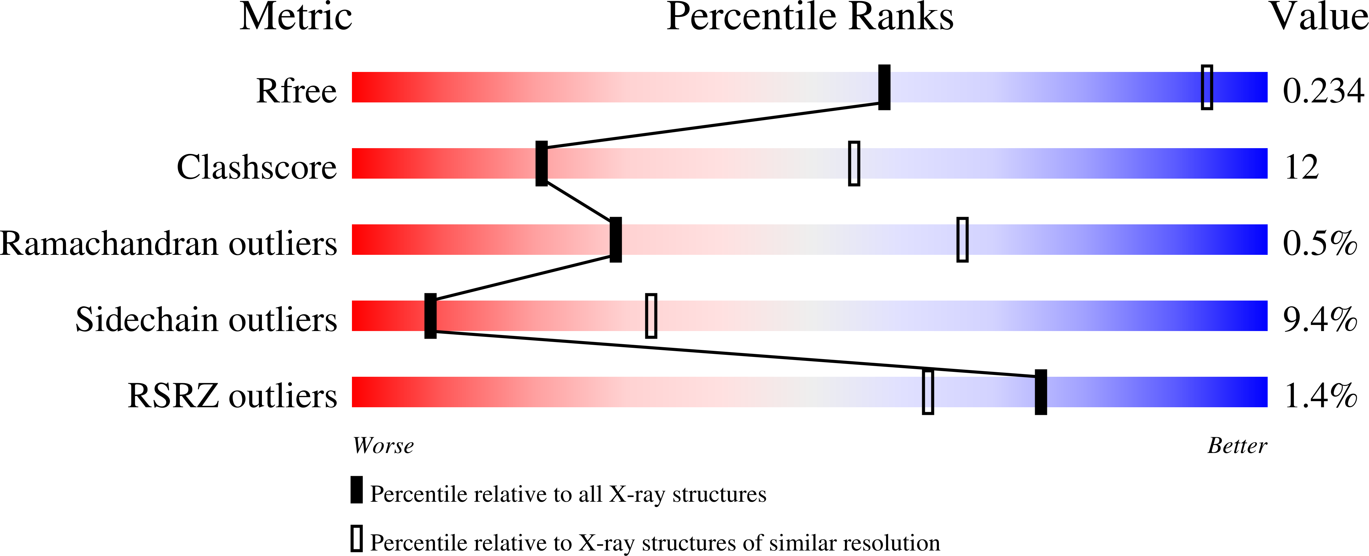
  • R-Value Free: 0.234 
  • R-Value Work: 0.225 
  • R-Value Observed: 0.225 

wwPDB Validation   3D Report Full Report


This is version 1.2 of the entry. See complete history


Literature

A novel co-crystal structure affords the design of gain-of-function lentiviral integrase mutants in the presence of modified PSIP1/LEDGF/p75

Hare, S.Shun, M.C.Gupta, S.S.Valkov, E.Engelman, A.Cherepanov, P.

(2009) PLoS Pathog 5: e1000259-e1000259

  • DOI: https://doi.org/10.1371/journal.ppat.1000259
  • Primary Citation of Related Structures:  
    3F9K

  • PubMed Abstract: 

    Lens epithelium derived growth factor (LEDGF), also known as PC4 and SFRS1 interacting protein 1 (PSIP1) and transcriptional co-activator p75, is the cellular binding partner of lentiviral integrase (IN) proteins. LEDGF accounts for the characteristic propensity of Lentivirus to integrate within active transcription units and is required for efficient viral replication. We now present a crystal structure containing the N-terminal and catalytic core domains (NTD and CCD) of HIV-2 IN in complex with the IN binding domain (IBD) of LEDGF. The structure extends the known IN-LEDGF interface, elucidating primarily charge-charge interactions between the NTD of IN and the IBD. A constellation of acidic residues on the NTD is characteristic of lentiviral INs, and mutations of the positively charged residues on the IBD severely affect interaction with all lentiviral INs tested. We show that the novel NTD-IBD contacts are critical for stimulation of concerted lentiviral DNA integration by LEDGF in vitro and for its function during the early steps of HIV-1 replication. Furthermore, the new structural details enabled us to engineer a mutant of HIV-1 IN that primarily functions only when presented with a complementary LEDGF mutant. These findings provide structural basis for the high affinity lentiviral IN-LEDGF interaction and pave the way for development of LEDGF-based targeting technologies for gene therapy.


  • Organizational Affiliation

    Division of Medicine, Imperial College London, St Mary's Campus, London, United Kingdom.


Macromolecules
Find similar proteins by:  (by identity cutoff)  |  3D Structure
Entity ID: 1
MoleculeChains Sequence LengthOrganismDetailsImage
Integrase210Human immunodeficiency virus type 2 (ISOLATE ROD)Mutation(s): 1 
Gene Names: gag-pol
UniProt
Find proteins for P04584 (Human immunodeficiency virus type 2 subtype A (isolate ROD))
Explore P04584 
Go to UniProtKB:  P04584
Entity Groups  
Sequence Clusters30% Identity50% Identity70% Identity90% Identity95% Identity100% Identity
UniProt GroupP04584
Sequence Annotations
Expand
  • Reference Sequence
Find similar proteins by:  (by identity cutoff)  |  3D Structure
Entity ID: 2
MoleculeChains Sequence LengthOrganismDetailsImage
PC4 and SFRS1-interacting protein95Homo sapiensMutation(s): 0 
Gene Names: PSIP1DFS70LEDGFPSIP2
UniProt & NIH Common Fund Data Resources
Find proteins for O75475 (Homo sapiens)
Explore O75475 
Go to UniProtKB:  O75475
PHAROS:  O75475
GTEx:  ENSG00000164985 
Entity Groups  
Sequence Clusters30% Identity50% Identity70% Identity90% Identity95% Identity100% Identity
UniProt GroupO75475
Sequence Annotations
Expand
  • Reference Sequence
Small Molecules
Ligands 2 Unique
IDChains Name / Formula / InChI Key2D Diagram3D Interactions
ZN
Query on ZN

Download Ideal Coordinates CCD File 
AB [auth Q]
AC [auth p]
CB [auth R]
CC [auth s]
EB [auth U]
AB [auth Q],
AC [auth p],
CB [auth R],
CC [auth s],
EB [auth U],
EC [auth t],
GB [auth V],
IB [auth Y],
KA [auth A],
KB [auth Z],
MA [auth B],
MB [auth c],
OA [auth E],
OB [auth d],
QA [auth F],
QB [auth g],
SA [auth I],
SB [auth h],
UA [auth J],
UB [auth k],
WA [auth M],
WB [auth l],
YA [auth N],
YB [auth o]
ZINC ION
Zn
PTFCDOFLOPIGGS-UHFFFAOYSA-N
MG
Query on MG

Download Ideal Coordinates CCD File 
BB [auth Q]
BC [auth p]
DB [auth R]
DC [auth s]
FB [auth U]
BB [auth Q],
BC [auth p],
DB [auth R],
DC [auth s],
FB [auth U],
FC [auth t],
HB [auth V],
JB [auth Y],
LA [auth A],
LB [auth Z],
NA [auth B],
NB [auth c],
PA [auth E],
PB [auth d],
RA [auth F],
RB [auth g],
TA [auth I],
TB [auth h],
VA [auth J],
VB [auth k],
XA [auth M],
XB [auth l],
ZA [auth N],
ZB [auth o]
MAGNESIUM ION
Mg
JLVVSXFLKOJNIY-UHFFFAOYSA-N
Experimental Data & Validation

Experimental Data

  • Method: X-RAY DIFFRACTION
  • Resolution: 3.20 Å
  • R-Value Free: 0.234 
  • R-Value Work: 0.225 
  • R-Value Observed: 0.225 
  • Space Group: P 21 21 21
Unit Cell:
Length ( Å )Angle ( ˚ )
a = 201.36α = 90
b = 202.5β = 90
c = 280.48γ = 90
Software Package:
Software NamePurpose
DNAdata collection
PHASERphasing
REFMACrefinement
MOSFLMdata reduction
SCALAdata scaling

Structure Validation

View Full Validation Report


Entry History 

Deposition Data

Revision History  (Full details and data files)

  • Version 1.0: 2009-01-20
    Type: Initial release
  • Version 1.1: 2011-07-13
    Changes: Derived calculations, Version format compliance
  • Version 1.2: 2023-11-01
    Changes: Data collection, Database references, Derived calculations, Refinement description