3EYI

The crystal structure of the second Z-DNA binding domain of human DAI (ZBP1) in complex with Z-DNA


Experimental Data Snapshot

  • Method: X-RAY DIFFRACTION
  • Resolution: 1.45 Å
  • R-Value Free: 0.194 
  • R-Value Work: 0.153 
  • R-Value Observed: 0.157 

wwPDB Validation   3D Report Full Report


This is version 1.2 of the entry. See complete history


Literature

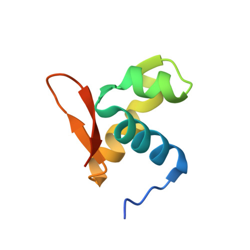
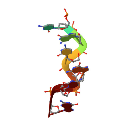
The crystal structure of the second Z-DNA binding domain of human DAI (ZBP1) in complex with Z-DNA reveals an unusual binding mode to Z-DNA.

Ha, S.C.Kim, D.Hwang, H.Y.Rich, A.Kim, Y.G.Kim, K.K.

(2008) Proc Natl Acad Sci U S A 105: 20671-20676

  • DOI: https://doi.org/10.1073/pnas.0810463106
  • Primary Citation of Related Structures:  
    3EYI

  • PubMed Abstract: 

    Mammalian DAI (DNA-dependent activator of IFN-regulatory factors), an activator of the innate immune response, senses cytosolic DNA by using 2 N-terminal Z-DNA binding domains (ZBDs) and a third putative DNA binding domain located next to the second ZBD. Compared with other previously known ZBDs, the second ZBD of human DAI (hZbeta(DAI)) shows significant variation in the sequence of the residues that are essential for DNA binding. In this article, the crystal structure of the hZbeta(DAI)/Z-DNA complex reveals that hZbeta(DAI) has a similar fold to that of other ZBDs, but adopts an unusual binding mode for recognition of Z-DNA. A residue in the first beta-strand rather than residues in the beta-loop contributes to DNA binding, and part of the (alpha3) recognition helix adopts a 3(10) helix conformation. The role of each residue that makes contact with DNA was confirmed by mutational analysis. The 2 ZBDs of DAI can together bind to DNA and both are necessary for full B-to-Z conversion. It is possible that binding 2 DAIs to 1 dsDNA brings about dimerization of DAI that might facilitate DNA-mediated innate immune activation.


  • Organizational Affiliation

    Department of Molecular Cell Biology, Samsung Biomedical Research Institute, Sungkyunkwan University School of Medicine, Suwon 440-746, Korea.


Macromolecules

Find similar proteins by:  (by identity cutoff)  |  3D Structure
Entity ID: 1
MoleculeChains Sequence LengthOrganismDetailsImage
Z-DNA-binding protein 1
A, B
72Homo sapiensMutation(s): 0 
Gene Names: C20orf183DLM1ZBP1ZBP1/DLM1
UniProt & NIH Common Fund Data Resources
Find proteins for Q9H171 (Homo sapiens)
Explore Q9H171 
Go to UniProtKB:  Q9H171
PHAROS:  Q9H171
GTEx:  ENSG00000124256 
Entity Groups  
Sequence Clusters30% Identity50% Identity70% Identity90% Identity95% Identity100% Identity
UniProt GroupQ9H171
Sequence Annotations
Expand
  • Reference Sequence

Find similar nucleic acids by:  Sequence   |   3D Structure  

Entity ID: 2
MoleculeChains LengthOrganismImage
5'-TCGCGCG-3'
C, D
7N/A
Sequence Annotations
Expand
  • Reference Sequence
Experimental Data & Validation

Experimental Data

  • Method: X-RAY DIFFRACTION
  • Resolution: 1.45 Å
  • R-Value Free: 0.194 
  • R-Value Work: 0.153 
  • R-Value Observed: 0.157 
  • Space Group: P 21 21 21
Unit Cell:
Length ( Å )Angle ( ˚ )
a = 29.53α = 90
b = 58.249β = 90
c = 88.609γ = 90
Software Package:
Software NamePurpose
MAR345dtbdata collection
autoSHARPphasing
REFMACrefinement
HKL-2000data reduction
SCALEPACKdata scaling

Structure Validation

View Full Validation Report


Entry History 

Deposition Data

Revision History  (Full details and data files)

  • Version 1.0: 2009-01-13
    Type: Initial release
  • Version 1.1: 2011-07-13
    Changes: Version format compliance
  • Version 1.2: 2023-12-27
    Changes: Data collection, Database references