3D8A

Co-crystal structure of TraM-TraD complex.


Experimental Data Snapshot

  • Method: X-RAY DIFFRACTION
  • Resolution: 2.55 Å
  • R-Value Free: 0.257 
  • R-Value Work: 0.218 
  • R-Value Observed: 0.220 

wwPDB Validation   3D Report Full Report


This is version 1.3 of the entry. See complete history


Literature

Structural basis of specific TraD-TraM recognition during F plasmid-mediated bacterial conjugation.

Lu, J.Wong, J.J.Edwards, R.A.Manchak, J.Frost, L.S.Glover, J.N.

(2008) Mol Microbiol 70: 89-99

  • DOI: https://doi.org/10.1111/j.1365-2958.2008.06391.x
  • Primary Citation of Related Structures:  
    3D8A

  • PubMed Abstract: 

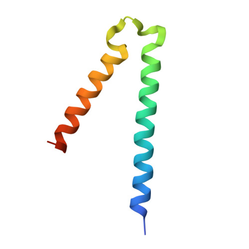
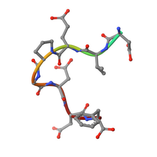
    F plasmid-mediated bacterial conjugation requires interactions between a relaxosome component, TraM, and the coupling protein TraD, a hexameric ring ATPase that forms the cytoplasmic face of the conjugative pore. Here we present the crystal structure of the C-terminal tail of TraD bound to the TraM tetramerization domain, the first structural evidence of relaxosome-coupling protein interactions. The structure reveals the TraD C-terminal peptide bound to each of four symmetry-related grooves on the surface of the TraM tetramer. Extensive protein-protein interactions were observed between the two proteins. Mutational analysis indicates that these interactions are specific and required for efficient F conjugation in vivo. Our results suggest that specific interactions between the C-terminal tail of TraD and the TraM tetramerization domain might lead to more generalized interactions that stabilize the relaxosome-coupling protein complex in preparation for conjugative DNA transfer.


  • Organizational Affiliation

    Departments of Biochemistry, University of Alberta, Edmonton, Alberta, Canada.


Macromolecules
Find similar proteins by:  (by identity cutoff)  |  3D Structure
Entity ID: 1
MoleculeChains Sequence LengthOrganismDetailsImage
Relaxosome protein TraM
A, B, C, D, E
A, B, C, D, E, F, G, H
70Escherichia coli K-12Mutation(s): 0 
Gene Names: traMECOK12F071
UniProt
Find proteins for P10026 (Escherichia coli (strain K12))
Explore P10026 
Go to UniProtKB:  P10026
Entity Groups  
Sequence Clusters30% Identity50% Identity70% Identity90% Identity95% Identity100% Identity
UniProt GroupP10026
Sequence Annotations
Expand
  • Reference Sequence

Find similar proteins by:  Sequence   |   3D Structure  

Entity ID: 2
MoleculeChains Sequence LengthOrganismDetailsImage
Protein traD10Escherichia coli K-12Mutation(s): 0 
Gene Names: traDECOK12F102
UniProt
Find proteins for P09130 (Escherichia coli (strain K12))
Explore P09130 
Go to UniProtKB:  P09130
Entity Groups  
Sequence Clusters30% Identity50% Identity70% Identity90% Identity95% Identity100% Identity
UniProt GroupP09130
Sequence Annotations
Expand
  • Reference Sequence
Experimental Data & Validation

Experimental Data

  • Method: X-RAY DIFFRACTION
  • Resolution: 2.55 Å
  • R-Value Free: 0.257 
  • R-Value Work: 0.218 
  • R-Value Observed: 0.220 
  • Space Group: H 3
Unit Cell:
Length ( Å )Angle ( ˚ )
a = 142.246α = 90
b = 142.246β = 90
c = 70.95γ = 120
Software Package:
Software NamePurpose
REFMACrefinement
Blu-Icedata collection
DENZOdata reduction
SCALEPACKdata scaling
MOLREPphasing

Structure Validation

View Full Validation Report


Entry History 

Deposition Data

Revision History  (Full details and data files)

  • Version 1.0: 2008-09-09
    Type: Initial release
  • Version 1.1: 2011-07-13
    Changes: Advisory, Version format compliance
  • Version 1.2: 2017-06-28
    Changes: Database references, Source and taxonomy, Structure summary
  • Version 1.3: 2023-08-30
    Changes: Data collection, Database references, Refinement description