


X-ray crystal structure of homoserine dehydrogenase from Thermoplasma volcanium.
Osipiuk, J., Hendricks, R., Abdullah, J., Joachimiak, A.To be published.
Experimental Data Snapshot
wwPDB Validation 3D Report Full Report

Entity ID: 1 | |||||
|---|---|---|---|---|---|
| Molecule | Chains | Sequence Length | Organism | Details | Image |
| Homoserine dehydrogenase | 331 | Thermoplasma volcanium GSS1 | Mutation(s): 0 Gene Names: TVG0375766, TV0389 | 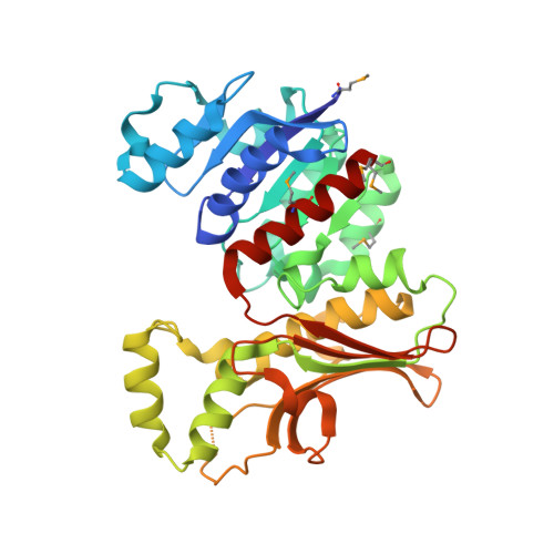 | |
UniProt | |||||
Find proteins for Q97BR6 (Thermoplasma volcanium (strain ATCC 51530 / DSM 4299 / JCM 9571 / NBRC 15438 / GSS1)) Explore Q97BR6 Go to UniProtKB: Q97BR6 | |||||
Entity Groups | |||||
| Sequence Clusters | 30% Identity50% Identity70% Identity90% Identity95% Identity100% Identity | ||||
| UniProt Group | Q97BR6 | ||||
Sequence AnnotationsExpand | |||||
| |||||
| Ligands 1 Unique | |||||
|---|---|---|---|---|---|
| ID | Chains | Name / Formula / InChI Key | 2D Diagram | 3D Interactions | |
| SO4 Query on SO4 | B [auth A], C [auth A], D [auth A], E [auth A] | SULFATE ION O4 S QAOWNCQODCNURD-UHFFFAOYSA-L |  | ||
| Modified Residues 1 Unique | |||||
|---|---|---|---|---|---|
| ID | Chains | Type | Formula | 2D Diagram | Parent |
| MSE Query on MSE | A | L-PEPTIDE LINKING | C5 H11 N O2 Se |  | MET |
| Length ( Å ) | Angle ( ˚ ) |
|---|---|
| a = 108.404 | α = 90 |
| b = 108.404 | β = 90 |
| c = 60.458 | γ = 120 |
| Software Name | Purpose |
|---|---|
| DENZO | data reduction |
| SCALEPACK | data scaling |
| REFMAC | refinement |
| PDB_EXTRACT | data extraction |
| SBC-Collect | data collection |
| HKL-3000 | data reduction |
| SHELXD | phasing |
| MLPHARE | phasing |
| DM | phasing |
| SOLVE | phasing |
| RESOLVE | phasing |
| HKL-3000 | phasing |
RCSB PDB (citation) is hosted by
RCSB PDB is a member of the

