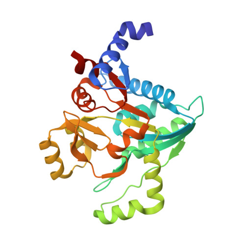
Crystal structures of the effector-binding domain of repressor Central glycolytic gene Regulator from Bacillus subtilis reveal ligand-induced structural changes upon binding of several glycolytic intermediates.
Rezacova, P., Kozisek, M., Moy, S.F., Sieglova, I., Joachimiak, A., Machius, M., Otwinowski, Z.(2008) Mol Microbiol 69: 895-910
- PubMed: 18554327
- DOI: https://doi.org/10.1111/j.1365-2958.2008.06318.x
- Primary Citation of Related Structures:
2OKG, 3BXE, 3BXF, 3BXG, 3BXH - PubMed Abstract:
Expression of genes in the gapA operon encoding five enzymes for triose phosphate interconversion in Bacillus subtilis is negatively regulated by the Central glycolytic genes Regulator (CggR). CggR belongs to the large SorC/DeoR family of prokaryotic transcriptional regulators, characterized by an N-terminal DNA-binding domain and a large C-terminal effector-binding domain. When no glucose is present in growth media, CggR binds to its target DNA sequence and blocks the transcription of genes in the gapA operon. In the presence of glucose, binding of the known effector molecule fructose-1,6-bisphosphate abolishes this interaction. We have identified dihydroxyacetone phosphate, glucose-6-phosphate and fructose-6-phosphate as additional CggR ligands that can bind to the effector-binding site. Crystal structures of C-CggR, the C-terminal effector-binding domain of CggR, both unliganded as well as in complex with the four ligands at resolutions between 1.65 and 1.80 A reveal unique ligand-specific structural changes in the binding site that affect the dimer interface. Binding affinities of these ligands were determined by isothermal titration calorimetry. Chemical cross-linking shows that CggR oligomerization is mediated through its effector-binding domain, and that binding of the different ligands differentially affects the distribution of oligomers. Electrophoretic mobility shift assays (EMSAs) confirmed a destabilizing effect of fructose-1,6-bisphosphate on the CggR/DNA complex, and also showed similar effects for dihydroxyacetone phosphate. Our results suggest that CggR stability and function may be modulated by various effectors in a complex fashion.
Organizational Affiliation:
Department of Biochemistry, The University of Texas Southwestern Medical Center at Dallas, Dallas, TX, USA. rezacova@uochb.cas.cz