3BWE

Crystal structure of aggregated form of DJ1


Experimental Data Snapshot

  • Method: X-RAY DIFFRACTION
  • Resolution: 2.40 Å
  • R-Value Free: 0.282 
  • R-Value Work: 0.232 
  • R-Value Observed: 0.232 

wwPDB Validation   3D Report Full Report


This is version 1.3 of the entry. See complete history


Literature

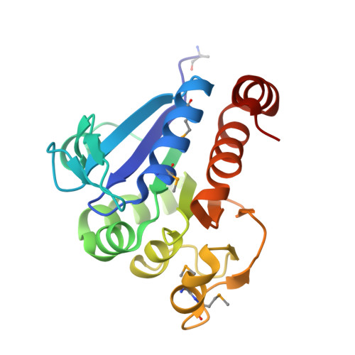
Crystal structure of filamentous aggregates of human DJ-1 formed in an inorganic phosphate-dependent manner.

Cha, S.S.Jung, H.I.Jeon, H.An, Y.J.Kim, I.K.Yun, S.Ahn, H.J.Chung, K.C.Lee, S.H.Suh, P.G.Kang, S.O.

(2008) J Biol Chem 283: 34069-34075

  • DOI: https://doi.org/10.1074/jbc.M804243200
  • Primary Citation of Related Structures:  
    3BWE

  • PubMed Abstract: 

    Mutations in the DJ-1 gene have been implicated in the autosomal recessive early onset parkinsonism. DJ-1 is a soluble dimeric protein with critical roles in response to oxidative stress and in neuronal maintenance. However, several lines of evidence suggest the existence of a nonfunctional aggregated form of DJ-1 in the brain of patients with some neurodegenerative diseases. Here, we show that inorganic phosphate, an important anion that exhibits elevated levels in patients with Parkinson disease, transforms DJ-1 into filamentous aggregates. According to the 2.4-A crystal structure, DJ-1 dimers are linearly stacked through P(i)-mediated interactions to form protofilaments, which are then bundled into a filamentous assembly.


  • Organizational Affiliation

    Marine and Extreme Genome Research Center, Korea Ocean Research & Development Institute, Ansan 426-744, Republic of Korea. chajung@kordi.re.kr


Macromolecules
Find similar proteins by:  (by identity cutoff)  |  3D Structure
Entity ID: 1
MoleculeChains Sequence LengthOrganismDetailsImage
Protein DJ-1
A, B, C, D, E
A, B, C, D, E, F, G
189Homo sapiensMutation(s): 0 
Gene Names: PARK7
UniProt & NIH Common Fund Data Resources
Find proteins for Q99497 (Homo sapiens)
Explore Q99497 
Go to UniProtKB:  Q99497
PHAROS:  Q99497
GTEx:  ENSG00000116288 
Entity Groups  
Sequence Clusters30% Identity50% Identity70% Identity90% Identity95% Identity100% Identity
UniProt GroupQ99497
Sequence Annotations
Expand
  • Reference Sequence
Experimental Data & Validation

Experimental Data

  • Method: X-RAY DIFFRACTION
  • Resolution: 2.40 Å
  • R-Value Free: 0.282 
  • R-Value Work: 0.232 
  • R-Value Observed: 0.232 
  • Space Group: C 1 2 1
Unit Cell:
Length ( Å )Angle ( ˚ )
a = 193.616α = 90
b = 102.582β = 113.69
c = 85.535γ = 90
Software Package:
Software NamePurpose
ADSCdata collection
AMoREphasing
CNSrefinement
HKL-2000data reduction
HKL-2000data scaling

Structure Validation

View Full Validation Report


Entry History 

Deposition Data

  • Released Date: 2008-10-14 
  • Deposition Author(s): Cha, S.S.

Revision History  (Full details and data files)

  • Version 1.0: 2008-10-14
    Type: Initial release
  • Version 1.1: 2011-07-13
    Changes: Version format compliance
  • Version 1.2: 2023-11-01
    Changes: Data collection, Database references, Derived calculations, Refinement description
  • Version 1.3: 2023-11-15
    Changes: Data collection