3BBD

M. jannaschii Nep1 complexed with S-adenosyl-homocysteine


Experimental Data Snapshot

  • Method: X-RAY DIFFRACTION
  • Resolution: 2.15 Å
  • R-Value Free: 0.221 
  • R-Value Work: 0.180 
  • R-Value Observed: 0.182 

wwPDB Validation   3D Report Full Report


Ligand Structure Quality Assessment 


This is version 1.1 of the entry. See complete history


Literature

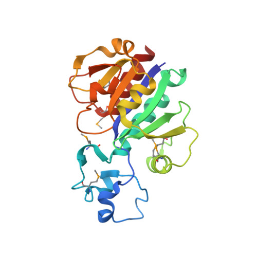
The crystal structure of Nep1 reveals an extended SPOUT-class methyltransferase fold and a pre-organized SAM-binding site.

Taylor, A.B.Meyer, B.Leal, B.Z.Kotter, P.Schirf, V.Demeler, B.Hart, P.J.Entian, K.D.Wohnert, J.

(2008) Nucleic Acids Res 36: 1542-1554

  • DOI: https://doi.org/10.1093/nar/gkm1172
  • Primary Citation of Related Structures:  
    3BBD, 3BBE, 3BBH

  • PubMed Abstract: 

    Ribosome biogenesis in eukaryotes requires the participation of a large number of ribosome assembly factors. The highly conserved eukaryotic nucleolar protein Nep1 has an essential but unknown function in 18S rRNA processing and ribosome biogenesis. In Saccharomyces cerevisiae the malfunction of a temperature-sensitive Nep1 protein (nep1-1(ts)) was suppressed by the addition of S-adenosylmethionine (SAM). This suggests the participation of Nep1 in a methyltransferase reaction during ribosome biogenesis. In addition, yeast Nep1 binds to a 6-nt RNA-binding motif also found in 18S rRNA and facilitates the incorporation of ribosomal protein Rps19 during the formation of pre-ribosomes. Here, we present the X-ray structure of the Nep1 homolog from the archaebacterium Methanocaldococcus jannaschii in its free form (2.2 A resolution) and bound to the S-adenosylmethionine analog S-adenosylhomocysteine (SAH, 2.15 A resolution) and the antibiotic and general methyltransferase inhibitor sinefungin (2.25 A resolution). The structure reveals a fold which is very similar to the conserved core fold of the SPOUT-class methyltransferases but contains a novel extension of this common core fold. SAH and sinefungin bind to Nep1 at a preformed binding site that is topologically equivalent to the cofactor-binding site in other SPOUT-class methyltransferases. Therefore, our structures together with previous genetic data suggest that Nep1 is a genuine rRNA methyltransferase.


  • Organizational Affiliation

    Department of Biochemistry, X-ray Crystallography Core Laboratory, The University of Texas Health Science Center San Antonio, San Antonio, TX-78229, USA.


Macromolecules
Find similar proteins by:  (by identity cutoff)  |  3D Structure
Entity ID: 1
MoleculeChains Sequence LengthOrganismDetailsImage
Ribosome biogenesis protein NEP1-like
A, B
205Methanocaldococcus jannaschiiMutation(s): 0 
Gene Names: Nep1
UniProt
Find proteins for Q57977 (Methanocaldococcus jannaschii (strain ATCC 43067 / DSM 2661 / JAL-1 / JCM 10045 / NBRC 100440))
Explore Q57977 
Go to UniProtKB:  Q57977
Entity Groups  
Sequence Clusters30% Identity50% Identity70% Identity90% Identity95% Identity100% Identity
UniProt GroupQ57977
Sequence Annotations
Expand
  • Reference Sequence
Small Molecules
Ligands 2 Unique
IDChains Name / Formula / InChI Key2D Diagram3D Interactions
SAH
Query on SAH

Download Ideal Coordinates CCD File 
C [auth A],
H [auth B]
S-ADENOSYL-L-HOMOCYSTEINE
C14 H20 N6 O5 S
ZJUKTBDSGOFHSH-WFMPWKQPSA-N
GOL
Query on GOL

Download Ideal Coordinates CCD File 
D [auth A]
E [auth A]
F [auth A]
G [auth A]
I [auth B]
D [auth A],
E [auth A],
F [auth A],
G [auth A],
I [auth B],
J [auth B],
K [auth B]
GLYCEROL
C3 H8 O3
PEDCQBHIVMGVHV-UHFFFAOYSA-N
Modified Residues  1 Unique
IDChains TypeFormula2D DiagramParent
MSE
Query on MSE
A, B
L-PEPTIDE LINKINGC5 H11 N O2 SeMET
Experimental Data & Validation

Experimental Data

  • Method: X-RAY DIFFRACTION
  • Resolution: 2.15 Å
  • R-Value Free: 0.221 
  • R-Value Work: 0.180 
  • R-Value Observed: 0.182 
  • Space Group: P 21 21 21
Unit Cell:
Length ( Å )Angle ( ˚ )
a = 84.894α = 90
b = 91.121β = 90
c = 93.713γ = 90
Software Package:
Software NamePurpose
PHENIXrefinement
ADSCdata collection
HKL-2000data reduction
HKL-2000data scaling
SHARPphasing

Structure Validation

View Full Validation Report


Ligand Structure Quality Assessment 


Entry History 

Deposition Data

Revision History  (Full details and data files)

  • Version 1.0: 2008-02-05
    Type: Initial release
  • Version 1.1: 2011-07-13
    Changes: Non-polymer description, Version format compliance