3ASV

The Closed form of serine dehydrogenase complexed with NADP+


Experimental Data Snapshot

  • Method: X-RAY DIFFRACTION
  • Resolution: 2.70 Å
  • R-Value Free: 0.273 
  • R-Value Work: 0.192 

wwPDB Validation   3D Report Full Report


Ligand Structure Quality Assessment 


This is version 1.1 of the entry. See complete history


Literature

Crystal structure of serine dehydrogenase from Escherichia coli: important role of the C-terminal region for closed-complex formation.

Yamazawa, R.Nakajima, Y.Mushiake, K.Yoshimoto, T.Ito, K.

(2011) J Biochem 149: 701-712

  • DOI: https://doi.org/10.1093/jb/mvr024
  • Primary Citation of Related Structures:  
    3ASU, 3ASV

  • PubMed Abstract: 

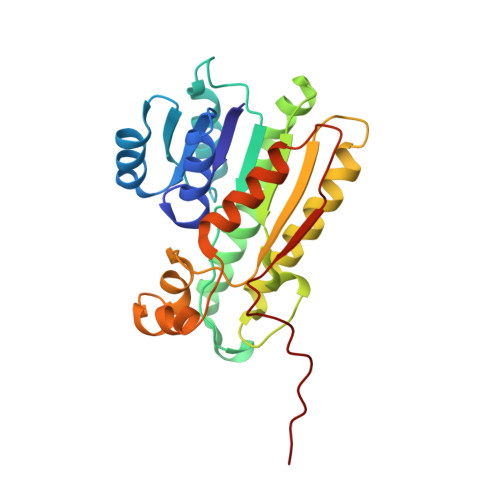
    Serine dehydrogenase from Escherichia coli is a homotetrameric enzyme belonging to the short-chain dehydrogenase/reductase (SDR) family. This enzyme catalyses the NADP(+)-dependent oxidation of serine to 2-aminomalonate semialdehyde. The enzyme shows a stereospecificity for β-(3S)-hydroxy acid as a substrate; however, no stereospecificity was observed at the α-carbon. The structures of the ligand-free SerDH and SerDH-NADP(+)-phosphate complex were determined at 1.9 and 2.7 Å resolutions, respectively. The overall structure, including the catalytic tetrad of Asn106, Ser134, Tyr147 and Lys151, shows obvious relationships with other members of the SDR family. The structure of the substrate-binding loop and that of the C-terminal region were disordered in the ligand-free enzyme, whereas these structures were clearly defined in the SerDH-NADP(+) complex as a closed form. Interestingly, the C-terminal region was protruded from the main body and it formed an anti-parallel β-sheet with another C-terminal region on the subunit that is diagonally opposite to that in the tetramer. It is revealed that the C-terminal region possesses the important roles in substrate binding through the stabilization of the substrate-binding loop in the closed form complex. The roles of the C-terminal region along with those of the residues involved in substrate recognition were studied by site-directed mutagenesis.


  • Organizational Affiliation

    Graduate School of Biomedical Sciences, Nagasaki University, 1-14 Bunkyo-machi, Nagasaki, Japan.


Macromolecules
Find similar proteins by:  (by identity cutoff)  |  3D Structure
Entity ID: 1
MoleculeChains Sequence LengthOrganismDetailsImage
Short-chain dehydrogenase/reductase SDR
A, B, C, D, E
A, B, C, D, E, F
248Escherichia coliMutation(s): 0 
Gene Names: EcDH1_2106YdfG
Entity Groups  
Sequence Clusters30% Identity50% Identity70% Identity90% Identity95% Identity100% Identity
Sequence Annotations
Expand
  • Reference Sequence
Small Molecules
Ligands 2 Unique
IDChains Name / Formula / InChI Key2D Diagram3D Interactions
NAP
Query on NAP

Download Ideal Coordinates CCD File 
G [auth A]
I [auth B]
K [auth C]
M [auth D]
O [auth E]
G [auth A],
I [auth B],
K [auth C],
M [auth D],
O [auth E],
Q [auth F]
NADP NICOTINAMIDE-ADENINE-DINUCLEOTIDE PHOSPHATE
C21 H28 N7 O17 P3
XJLXINKUBYWONI-NNYOXOHSSA-N
PO4
Query on PO4

Download Ideal Coordinates CCD File 
H [auth A]
J [auth B]
L [auth C]
N [auth D]
P [auth E]
H [auth A],
J [auth B],
L [auth C],
N [auth D],
P [auth E],
R [auth F]
PHOSPHATE ION
O4 P
NBIIXXVUZAFLBC-UHFFFAOYSA-K
Experimental Data & Validation

Experimental Data

  • Method: X-RAY DIFFRACTION
  • Resolution: 2.70 Å
  • R-Value Free: 0.273 
  • R-Value Work: 0.192 
  • Space Group: P 64
Unit Cell:
Length ( Å )Angle ( ˚ )
a = 113.859α = 90
b = 113.859β = 90
c = 197.54γ = 120
Software Package:
Software NamePurpose
SCALEPACKdata scaling
PHASERphasing
CNSrefinement
PDB_EXTRACTdata extraction
CrystalCleardata collection
HKL-2000data reduction
HKL-2000data scaling

Structure Validation

View Full Validation Report


Ligand Structure Quality Assessment 


Entry History 

Deposition Data

Revision History  (Full details and data files)

  • Version 1.0: 2011-10-12
    Type: Initial release
  • Version 1.1: 2023-11-01
    Changes: Data collection, Database references, Derived calculations, Refinement description