3AG0

Crystal structure of carbonmonoxy human cytoglobin


Experimental Data Snapshot

  • Method: X-RAY DIFFRACTION
  • Resolution: 2.60 Å
  • R-Value Free: 0.248 
  • R-Value Work: 0.215 
  • R-Value Observed: 0.215 

wwPDB Validation   3D Report Full Report


Ligand Structure Quality Assessment 


This is version 1.3 of the entry. See complete history


Literature

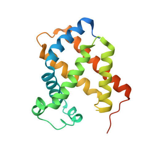
Crystal structure of the carbon monoxide complex of human cytoglobin

Makino, M.Sawai, H.Shiro, Y.Sugimoto, H.

(2011) Proteins 79: 1143-1153

  • DOI: https://doi.org/10.1002/prot.22950
  • Primary Citation of Related Structures:  
    3AG0

  • PubMed Abstract: 

    Cytoglobin (Cgb) is a vertebrate heme-containing globin-protein expressed in a broad range of mammalian tissues. Unlike myoglobin, Cgb displays a hexa-coordinated (bis-hystidyl) heme iron atom, having the heme distal His81(E7) residue as the endogenous sixth ligand. In the present study, we crystallized human Cgb in the presence of a reductant Na₂S₂O₄ under a carbon monoxide (CO) atmosphere, and determined the crystal structure at 2.6 A resolution. The CO ligand occupies the sixth axial position of the heme ferrous iron. Eventually, the imidazole group of His81(E7) is expelled from the sixth position and swings out of the distal heme pocket. The flipping motion of the His81 imidazole group accompanies structural readjustments of some residues (Gln62, Phe63, Gln72, and Ser75) in both the CD-corner and D-helix regions of Cgb. On the other hand, no significant structural changes were observed in other Cgb regions, for example, on the proximal side. These structural alterations that occurred as a result of exogenous ligand (CO) binding are clearly different from those observed in other vertebrate hexa-coordinated globins (mouse neuroglobin, Drosophila melanogaster hemoglobin) and penta-coordinated sperm whale myoglobin. The present study provides the structural basis for further discussion of the unique ligand-binding properties of Cgb.


  • Organizational Affiliation

    RIKEN SPring-8 Center, Sayo, Hyogo 679-5148, Japan.


Macromolecules
Find similar proteins by:  (by identity cutoff)  |  3D Structure
Entity ID: 1
MoleculeChains Sequence LengthOrganismDetailsImage
Cytoglobin192Homo sapiensMutation(s): 0 
Gene Names: CYGBSTAP
UniProt & NIH Common Fund Data Resources
Find proteins for Q8WWM9 (Homo sapiens)
Explore Q8WWM9 
Go to UniProtKB:  Q8WWM9
PHAROS:  Q8WWM9
GTEx:  ENSG00000161544 
Entity Groups  
Sequence Clusters30% Identity50% Identity70% Identity90% Identity95% Identity100% Identity
UniProt GroupQ8WWM9
Sequence Annotations
Expand
  • Reference Sequence
Small Molecules
Ligands 2 Unique
IDChains Name / Formula / InChI Key2D Diagram3D Interactions
HEM
Query on HEM

Download Ideal Coordinates CCD File 
B [auth A]PROTOPORPHYRIN IX CONTAINING FE
C34 H32 Fe N4 O4
KABFMIBPWCXCRK-RGGAHWMASA-L
CMO
Query on CMO

Download Ideal Coordinates CCD File 
C [auth A]CARBON MONOXIDE
C O
UGFAIRIUMAVXCW-UHFFFAOYSA-N
Experimental Data & Validation

Experimental Data

  • Method: X-RAY DIFFRACTION
  • Resolution: 2.60 Å
  • R-Value Free: 0.248 
  • R-Value Work: 0.215 
  • R-Value Observed: 0.215 
  • Space Group: P 21 21 21
Unit Cell:
Length ( Å )Angle ( ˚ )
a = 36.198α = 90
b = 43.977β = 90
c = 94.019γ = 90
Software Package:
Software NamePurpose
XSCALEdata scaling
MOLREPphasing
CNSrefinement
PDB_EXTRACTdata extraction
BSSdata collection
DENZOdata reduction
SCALEPACKdata scaling

Structure Validation

View Full Validation Report


Ligand Structure Quality Assessment 


Entry History 

Deposition Data

Revision History  (Full details and data files)

  • Version 1.0: 2011-02-02
    Type: Initial release
  • Version 1.1: 2011-07-13
    Changes: Version format compliance
  • Version 1.2: 2023-07-12
    Changes: Data collection, Database references, Derived calculations, Structure summary
  • Version 1.3: 2023-11-08
    Changes: Data collection, Database references, Refinement description