


Crystal structure of Entamoeba histolytica methionine gamma-lyase 1
Karaki, T., Sato, D., Shimizu, A., Nozaki, T., Harada, S.To be published.
Experimental Data Snapshot
Entity ID: 1 | |||||
|---|---|---|---|---|---|
| Molecule | Chains | Sequence Length | Organism | Details | Image |
| Methionine gamma-lyase | 389 | Entamoeba histolytica | Mutation(s): 0 Gene Names: metg EC: 4.4.1.11 | 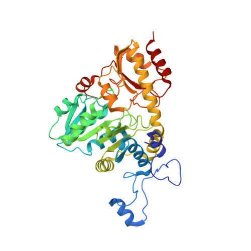 | |
UniProt | |||||
Find proteins for Q86D28 (Entamoeba histolytica) Explore Q86D28 Go to UniProtKB: Q86D28 | |||||
Entity Groups | |||||
| Sequence Clusters | 30% Identity50% Identity70% Identity90% Identity95% Identity100% Identity | ||||
| UniProt Group | Q86D28 | ||||
Sequence AnnotationsExpand | |||||
| |||||
Entity ID: 2 | |||||
|---|---|---|---|---|---|
| Molecule | Chains | Sequence Length | Organism | Details | Image |
| Methionine gamma-lyase | 389 | Entamoeba histolytica | Mutation(s): 0 Gene Names: metg EC: 4.4.1.11 | 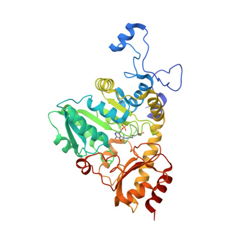 | |
UniProt | |||||
Find proteins for Q86D28 (Entamoeba histolytica) Explore Q86D28 Go to UniProtKB: Q86D28 | |||||
Entity Groups | |||||
| Sequence Clusters | 30% Identity50% Identity70% Identity90% Identity95% Identity100% Identity | ||||
| UniProt Group | Q86D28 | ||||
Sequence AnnotationsExpand | |||||
| |||||
| Ligands 4 Unique | |||||
|---|---|---|---|---|---|
| ID | Chains | Name / Formula / InChI Key | 2D Diagram | 3D Interactions | |
| AA5 Query on AA5 | E [auth A], H [auth B], M [auth D] | N-[(3-HYDROXY-2-METHYL-5-{[(TRIHYDROXYPHOSPHORANYL)OXY]METHYL}PYRIDIN-4-YL)METHYLENE]METHIONINE C13 H19 N2 O7 P S HPCOPQVFKQIVQF-DMSFABOWSA-N |  | ||
| MET Query on MET | J [auth C] | METHIONINE C5 H11 N O2 S FFEARJCKVFRZRR-BYPYZUCNSA-N |  | ||
| SO4 Query on SO4 | F [auth A], I [auth B], K [auth C], N [auth D] | SULFATE ION O4 S QAOWNCQODCNURD-UHFFFAOYSA-L |  | ||
| GOL Query on GOL | G [auth A], L [auth C] | GLYCEROL C3 H8 O3 PEDCQBHIVMGVHV-UHFFFAOYSA-N |  | ||
| Modified Residues 1 Unique | |||||
|---|---|---|---|---|---|
| ID | Chains | Type | Formula | 2D Diagram | Parent |
| LLP Query on LLP | C | L-PEPTIDE LINKING | C14 H22 N3 O7 P |  | LYS |
| Length ( Å ) | Angle ( ˚ ) |
|---|---|
| a = 98.738 | α = 90 |
| b = 85.359 | β = 101.92 |
| c = 113.664 | γ = 90 |
| Software Name | Purpose |
|---|---|
| DENZO | data reduction |
| SCALEPACK | data scaling |
| MOLREP | phasing |
| REFMAC | refinement |
| PDB_EXTRACT | data extraction |
| HKL-2000 | data collection |
| HKL-2000 | data reduction |
| HKL-2000 | data scaling |
RCSB PDB (citation) is hosted by
RCSB PDB is a member of the

