3A70

Crystal structure of Pseudomonas sp. MIS38 lipase in complex with diethyl phosphate


Experimental Data Snapshot

  • Method: X-RAY DIFFRACTION
  • Resolution: 2.15 Å
  • R-Value Free: 0.222 
  • R-Value Work: 0.170 
  • R-Value Observed: 0.173 

wwPDB Validation   3D Report Full Report


This is version 1.2 of the entry. See complete history


Literature

X-ray Crystallographic and MD Simulation Studies on the Mechanism of Interfacial Activation of a Family I.3 Lipase with Two Lids

Angkawidjaja, C.Matsumura, H.Koga, Y.Takano, K.Kanaya, S.

(2010) J Mol Biol 

  • DOI: https://doi.org/10.1016/j.jmb.2010.04.051
  • Primary Citation of Related Structures:  
    2ZVD, 3A6Z, 3A70

  • PubMed Abstract: 

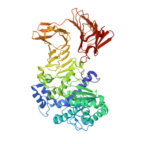
    The interfacial activation mechanism of family I.3 lipase from Pseudomonas sp. MIS38 (PML), which has two alpha-helical lids (lid1 and lid2), was investigated using a combination of X-ray crystallography and molecular dynamics (MD) simulation. The crystal structure of PML in an open conformation was determined at 2.1 A resolution in the presence of Ca(2+) and Triton X-100. Comparison of this structure with that in the closed conformation indicates that both lids greatly change their positions and lid1 is anchored by the calcium ion (Ca1) in the open conformation. This structure was not seriously changed even when the protein was dialyzed extensively against the Ca(2+)-free buffer containing Triton X-100 before crystallization, indicating that the open conformation is fairly stable unless a micellar substance is removed. The crystal structure of the PML derivative, in which the active site serine residue (Ser207) is diethylphosphorylated by soaking the crystal of PML in the open conformation in a solution containing diethyl p-nitrophenyl phosphate, was also determined. This structure greatly resembles that in the open conformation, indicating that PML structure in the open conformation represents that in the active form. MD simulation of PML in the open conformation in the absence of micelles showed that lid2 closes first, while lid1 maintains its open conformation. Likewise, MD simulation of PML in the closed conformation in the absence of Ca(2+) and in the presence of octane or trilaurin micelles showed that lid1 opens, while lid2 remains closed. These results suggest that Ca1 functions as a hook for stabilization of a fully opened conformation of lid1 and for initiation of subsequent opening of lid2.


  • Organizational Affiliation

    Department of Material and Life Science, Graduate School of Engineering, Osaka University, 2-1 Yamadaoka, Suita, Osaka 565-0871, Japan.


Macromolecules
Find similar proteins by:  (by identity cutoff)  |  3D Structure
Entity ID: 1
MoleculeChains Sequence LengthOrganismDetailsImage
LipaseA,
B [auth C]
617Pseudomonas sp. MIS38Mutation(s): 0 
EC: 3.1.1.3
UniProt
Find proteins for Q9RBY1 (Pseudomonas sp. MIS38)
Explore Q9RBY1 
Go to UniProtKB:  Q9RBY1
Entity Groups  
Sequence Clusters30% Identity50% Identity70% Identity90% Identity95% Identity100% Identity
UniProt GroupQ9RBY1
Sequence Annotations
Expand
  • Reference Sequence
Small Molecules
Ligands 4 Unique
IDChains Name / Formula / InChI Key2D Diagram3D Interactions
NPO
Query on NPO

Download Ideal Coordinates CCD File 
CA [auth C],
O [auth A]
P-NITROPHENOL
C6 H5 N O3
BTJIUGUIPKRLHP-UHFFFAOYSA-N
DEP
Query on DEP

Download Ideal Coordinates CCD File 
BA [auth C],
N [auth A]
DIETHYL PHOSPHONATE
C4 H11 O3 P
MJUJXFBTEFXVKU-UHFFFAOYSA-N
ACT
Query on ACT

Download Ideal Coordinates CCD File 
DA [auth C],
P [auth A]
ACETATE ION
C2 H3 O2
QTBSBXVTEAMEQO-UHFFFAOYSA-M
CA
Query on CA

Download Ideal Coordinates CCD File 
AA [auth C]
C [auth A]
D [auth A]
E [auth A]
F [auth A]
AA [auth C],
C [auth A],
D [auth A],
E [auth A],
F [auth A],
G [auth A],
H [auth A],
I [auth A],
J [auth A],
K [auth A],
L [auth A],
M [auth A],
Q [auth C],
R [auth C],
S [auth C],
T [auth C],
U [auth C],
V [auth C],
W [auth C],
X [auth C],
Y [auth C],
Z [auth C]
CALCIUM ION
Ca
BHPQYMZQTOCNFJ-UHFFFAOYSA-N
Experimental Data & Validation

Experimental Data

  • Method: X-RAY DIFFRACTION
  • Resolution: 2.15 Å
  • R-Value Free: 0.222 
  • R-Value Work: 0.170 
  • R-Value Observed: 0.173 
  • Space Group: P 65 2 2
Unit Cell:
Length ( Å )Angle ( ˚ )
a = 104.297α = 90
b = 104.297β = 90
c = 496.182γ = 120
Software Package:
Software NamePurpose
BL38B1data collection
MOLREPphasing
REFMACrefinement
HKL-2000data reduction
HKL-2000data scaling

Structure Validation

View Full Validation Report


Entry History 

Deposition Data

Revision History  (Full details and data files)

  • Version 1.0: 2010-05-26
    Type: Initial release
  • Version 1.1: 2011-07-13
    Changes: Version format compliance
  • Version 1.2: 2023-11-01
    Changes: Data collection, Database references, Derived calculations, Refinement description