3A1V

Crystal structue of the cytosolic domain of T. maritima FeoB iron iransporter in apo form


Experimental Data Snapshot

  • Method: X-RAY DIFFRACTION
  • Resolution: 2.40 Å
  • R-Value Free: 0.242 
  • R-Value Work: 0.203 
  • R-Value Observed: 0.205 

wwPDB Validation   3D Report Full Report


This is version 1.2 of the entry. See complete history


Literature

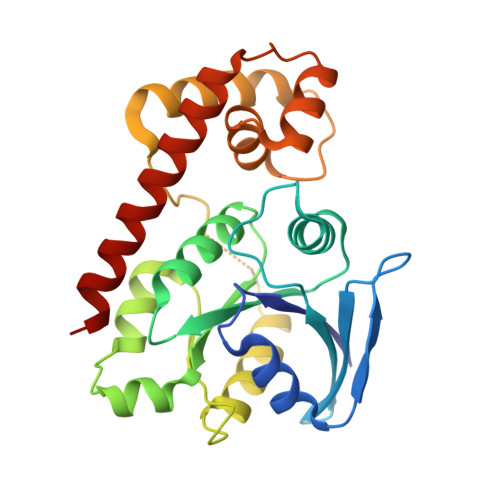
Structural basis of novel interactions between the small-GTPase and GDI-like domains in prokaryotic FeoB iron transporter

Hattori, M.Jin, Y.Nishimasu, H.Tanaka, Y.Mochizuki, M.Uchiumi, T.Ishitani, R.Ito, K.Nureki, O.

(2009) Structure 17: 1345-1355

  • DOI: https://doi.org/10.1016/j.str.2009.08.007
  • Primary Citation of Related Structures:  
    3A1S, 3A1T, 3A1U, 3A1V, 3A1W

  • PubMed Abstract: 

    The FeoB family proteins are widely distributed prokaryotic membrane proteins involved in Fe(2+) uptake. FeoB consists of N-terminal cytosolic and C-terminal transmembrane domains. The N-terminal region of the cytosolic domain is homologous to small GTPase (G) proteins and is considered to regulate Fe(2+) uptake. The spacer region connecting the G and TM domains reportedly functions as a GDP dissociation inhibitor (GDI)-like domain that stabilizes the GDP-binding state. However, the function of the G and GDI-like domains in iron uptake remains unclear. Here, we report the structural and functional analyses of the FeoB cytosolic domain from Thermotoga maritima. The structure-based mutational analysis indicated that the interaction between the G and GDI-like domains is important for both the GDI and Fe(2+) uptake activities. On the basis of these results, we propose a regulatory mechanism of Fe(2+) uptake.


  • Organizational Affiliation

    Department of Basic Medical Sciences, Institute of Medical Science, The University of Tokyo, Minato-ku, Tokyo, Japan.


Macromolecules
Find similar proteins by:  (by identity cutoff)  |  3D Structure
Entity ID: 1
MoleculeChains Sequence LengthOrganismDetailsImage
Iron(II) transport protein B
A, B
258Thermotoga maritimaMutation(s): 0 
Gene Names: TM0051
UniProt
Find proteins for Q9WXQ8 (Thermotoga maritima (strain ATCC 43589 / DSM 3109 / JCM 10099 / NBRC 100826 / MSB8))
Explore Q9WXQ8 
Go to UniProtKB:  Q9WXQ8
Entity Groups  
Sequence Clusters30% Identity50% Identity70% Identity90% Identity95% Identity100% Identity
UniProt GroupQ9WXQ8
Sequence Annotations
Expand
  • Reference Sequence
Small Molecules
Ligands 4 Unique
IDChains Name / Formula / InChI Key2D Diagram3D Interactions
EPE
Query on EPE

Download Ideal Coordinates CCD File 
E [auth A]4-(2-HYDROXYETHYL)-1-PIPERAZINE ETHANESULFONIC ACID
C8 H18 N2 O4 S
JKMHFZQWWAIEOD-UHFFFAOYSA-N
MRD
Query on MRD

Download Ideal Coordinates CCD File 
F [auth B](4R)-2-METHYLPENTANE-2,4-DIOL
C6 H14 O2
SVTBMSDMJJWYQN-RXMQYKEDSA-N
MPD
Query on MPD

Download Ideal Coordinates CCD File 
D [auth A],
G [auth B]
(4S)-2-METHYL-2,4-PENTANEDIOL
C6 H14 O2
SVTBMSDMJJWYQN-YFKPBYRVSA-N
SO4
Query on SO4

Download Ideal Coordinates CCD File 
C [auth A],
H [auth B]
SULFATE ION
O4 S
QAOWNCQODCNURD-UHFFFAOYSA-L
Experimental Data & Validation

Experimental Data

  • Method: X-RAY DIFFRACTION
  • Resolution: 2.40 Å
  • R-Value Free: 0.242 
  • R-Value Work: 0.203 
  • R-Value Observed: 0.205 
  • Space Group: P 21 21 21
Unit Cell:
Length ( Å )Angle ( ˚ )
a = 57.426α = 90
b = 81.556β = 90
c = 128.473γ = 90
Software Package:
Software NamePurpose
PHENIXrefinement

Structure Validation

View Full Validation Report


Entry History 

Deposition Data

Revision History  (Full details and data files)

  • Version 1.0: 2009-09-22
    Type: Initial release
  • Version 1.1: 2011-07-13
    Changes: Version format compliance
  • Version 1.2: 2023-11-01
    Changes: Data collection, Database references, Derived calculations, Refinement description