Theoretical and experimental studies of the conversion of chromopyrrolic acid to an antitumor derivative by cytochrome P450 StaP: the catalytic role of water molecules
Wang, Y., Chen, H., Makino, M., Shiro, Y., Nagano, S., Asamizu, S., Onaka, H., Shaik, S.(2009) J Am Chem Soc 131: 6748-6762
- PubMed: 19385626
- DOI: https://doi.org/10.1021/ja9003365
- Primary Citation of Related Structures:
3A1L - PubMed Abstract:
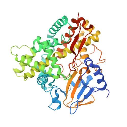
Chromopyrrolic acid (CPA) oxidation by cytochrome P450 StaP is a key process in the biosynthesis of antitumor drugs (Onaka, H.; Taniguchi, S.; Igarashi, Y.; Furumai, T. Biosci. Biotechnol. Biochem. 2003, 67, 127-138), which proceeds by an unusual C-C bond coupling. Additionally, because CPA is immobilized by a hydrogen-bonding array, it is prohibited from undergoing direct reaction with Compound I, the active species of P450. As such, the mechanism of P450 StaP poses a puzzle. In the present Article, we resolve this puzzle by combination of theory, using QM/MM calculations, and experiment, using crystallography and reactivity studies. Theory shows that the hydrogen-bonding machinery of the pocket deprotonates the carboxylic acid groups of CPA, while the nearby His(250) residue and the crystal waters, Wat(644) and Wat(789), assist the doubly deprotonated CPA to transfer electron density to Compound I; hence, CPA is activated toward proton-coupled electron transfer that sets the entire mechanism in motion. The ensuing mechanism involves a step of C-C bond formation coupled to a second electron transfer, four proton-transfer and tautomerization steps, and four steps where Wat(644) and Wat(789) move about and mediate these events. Experiments with the dichlorinated substrate, CCA, which expels Wat(644), show that the enzyme loses its activity. H250A and H250F mutations of P450 StaP show that His(250) is important, but in its absence Wat(644) and Wat(789) form a hydrogen-bonding diad that mediates the transformation. Thus, the water diad emerges as the minimal requisite element that endows StaP with function. This highlights the role of water molecules as biological catalysts that transform a P450 to a peroxidase-type (Derat, E.; Shaik, S. J. Am. Chem. Soc. 2006, 128, 13940-13949).
Organizational Affiliation:
The Institute of Chemistry and the Lise Meitner-Minerva Center for Computational Quantum Chemistry, Hebrew University of Jerusalem, Givat Ram Campus, 91904 Jerusalem, Israel.