386D

THREE-DIMENSIONAL STRUCTURE AND REACTIVITY OF A PHOTOCHEMICAL CLEAVAGE AGENT BOUND TO DNA


Experimental Data Snapshot

  • Method: X-RAY DIFFRACTION
  • Resolution: 1.80 Å
  • R-Value Free: 0.268 
  • R-Value Work: 0.196 
  • R-Value Observed: 0.196 

wwPDB Validation   3D Report Full Report


Ligand Structure Quality Assessment 


This is version 2.0 of the entry. See complete history


Literature

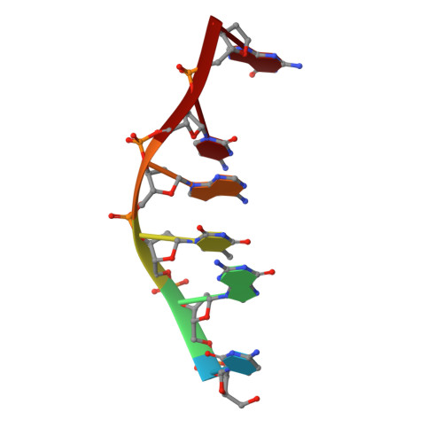
Three-Dimensional Structure and Reactivity of a Photochemical Cleavage Agent Bound to DNA

Gasper, S.M.Armitage, B.Shui, X.Hu, G.G.Yu, C.Schuster, G.Williams, L.D.

(1998) J Am Chem Soc 120: 12402-12409


Macromolecules

Find similar nucleic acids by:  Sequence   |   3D Structure  

Entity ID: 1
MoleculeChains LengthOrganismImage
DNA (5'-D(*CP*GP*TP*AP*CP*G)-3')6N/A
Sequence Annotations
Expand
  • Reference Sequence
Small Molecules
Ligands 1 Unique
IDChains Name / Formula / InChI Key2D Diagram3D Interactions
AQS
Query on AQS

Download Ideal Coordinates CCD File 
B [auth A]N,N-BIS(3-AMINOPROPYL)-2-ANTHRAQUINONESULFONAMIDE
C20 H25 N3 O4 S
HUHBNNVFLMYFIE-UHFFFAOYSA-P
Experimental Data & Validation

Experimental Data

  • Method: X-RAY DIFFRACTION
  • Resolution: 1.80 Å
  • R-Value Free: 0.268 
  • R-Value Work: 0.196 
  • R-Value Observed: 0.196 
  • Space Group: P 65 2 2
Unit Cell:
Length ( Å )Angle ( ˚ )
a = 31.02α = 90
b = 31.02β = 90
c = 64.91γ = 120
Software Package:
Software NamePurpose
MADmodel building
X-PLORrefinement
ADSCdata collection
MADphasing

Structure Validation

View Full Validation Report


Ligand Structure Quality Assessment 


Entry History 

Deposition Data

Revision History  (Full details and data files)

  • Version 1.0: 1998-03-12
    Type: Initial release
  • Version 1.1: 2008-05-22
    Changes: Version format compliance
  • Version 1.2: 2011-07-13
    Changes: Version format compliance
  • Version 2.0: 2024-02-21
    Changes: Atomic model, Data collection, Database references, Derived calculations