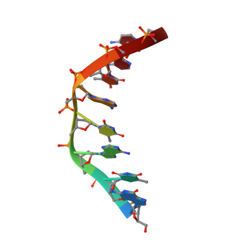
Structure of the side-by-side binding of distamycin to d(GTATATAC)2.
Mitra, S.N., Wahl, M.C., Sundaralingam, M.(1999) Acta Crystallogr D Biol Crystallogr 55: 602-609
- PubMed: 10089456
- DOI: https://doi.org/10.1107/s0907444998012475
- Primary Citation of Related Structures:
378D - PubMed Abstract:
The 2.40 A resolution crystal structure of a side-by-side binding of distamycin A molecules to a DNA octamer d(GTATATAC)2 with an extended alternating TA sequence has been determined. The unit-cell parameters are a = 29.55, b = 42.18, c = 43.38 A, beta = 96.56 degrees, space group P21, with two molecules in the asymmetric unit, in contrast to all previous side-by-side distamycin-DNA complexes which have only a single DNA strand and one drug molecule in the asymmetric unit. The structure was solved by the molecular-replacement method and refined to an R index of 21.0% using 3467 reflections [>/= 2sigma(F)]. The minor grooves of the DNA molecules bind two side-by-side antiparallel staggered distamycins spanning about five base pairs and virtually covering the entire length of the DNA. The octamer duplexes exhibit low-high alternations in the helical twist, sugar puckering and the C-O3' and O3'-P torsion angles, similar to the earlier side-by-side complexes containing inosine bases. The molecules are stacked one over the other along the ac diagonal in an infinite pseudo-continuous helical column with no lateral interactions.
Organizational Affiliation:
The Ohio State University, Biological Macromolecular Structure Center, Department of Chemistry, 012 Rightmire Hall, 1060 Carmack Road, Columbus, Ohio 43210-1002, USA.