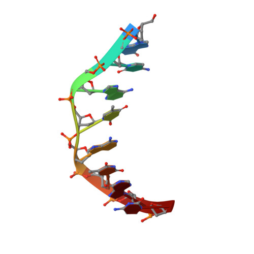
Crystal structures of the side-by-side binding of distamycin to AT-containing DNA octamers d(ICITACIC) and d(ICATATIC).
Chen, X., Ramakrishnan, B., Sundaralingam, M.(1997) J Mol Biol 267: 1157-1170
- PubMed: 9150404
- DOI: https://doi.org/10.1006/jmbi.1997.0941
- Primary Citation of Related Structures:
304D, 305D, 306D - PubMed Abstract:
To understand the recognition interactions between AT-containing alternating DNA and minor groove binding drugs, the crystal structures of the side-by-side binding of two distamycin molecules to the DNA octamers d(ICITACIC)2 and d(ICATATIC)2, referred to here as TA and ATAT, respectively, have been determined at 1.6 A and 2.2 A, respectively. Compared to the previous 2:1 all-IC d(ICICICIC)2-distamycin complex, the substitutions of the I x C base-pairs by the A x T base-pairs enable the interactions of the drug with its natural target to be studied. Both complexes assume side-by-side drug binding, isomorphous to the all IC counterpart in the tetragonal space group P4(1)22 (a = b = 28.03 A, c = 58.04 A and a = b = 27.86 A, c = 58.62 A, respectively). The ATAT complex also crystallized in a new polymorphic monoclinic space group C2 (a = 33.38 A, b = 25.33 A, c = 28.11 A and beta = 120.45 degrees) and was solved at 1.9 A resolution. The structures of the three double drug x DNA complexes are very similar, characterized by systematic hydrogen bonding and van der Waals interactions. Each drug hydrogen bonds with the bases of the proximal DNA strand only and stacks with the sugar moiety, while the side-by-side drugs themselves exhibit pyrrole ring-peptide stacking. The pyrrole-peptide interaction is crucial for the side-by-side binding mode of the distamycin/netropsin family of drugs. The purine-pyrimidine alternation is probably responsible for the striking alternation in the helical and backbone conformations. The structures are conserved between the pure IC complex and the AT substituted complexes but further details of the side-by-side binding to DNA are provided by the 1.6 A resolution structure of TA.
Organizational Affiliation:
Department of Chemistry, The Ohio State University, Columbus 43210, USA.