2ZRS

Crystal structure of Ca2+-bound form of des3-23ALG-2


Experimental Data Snapshot

  • Method: X-RAY DIFFRACTION
  • Resolution: 3.10 Å
  • R-Value Free: 0.280 
  • R-Value Work: 0.222 
  • R-Value Observed: 0.228 

wwPDB Validation   3D Report Full Report


This is version 1.3 of the entry. See complete history


Literature

Crystallization and X-ray diffraction analysis of N-terminally truncated human ALG-2

Suzuki, H.Kawasaki, M.Kakiuchi, T.Shibata, H.Wakatsuki, S.Maki, M.

(2008) Acta Crystallogr Sect F Struct Biol Cryst Commun 64: 974-977

  • DOI: https://doi.org/10.1107/S1744309108030297
  • Primary Citation of Related Structures:  
    2ZRS, 2ZRT

  • PubMed Abstract: 

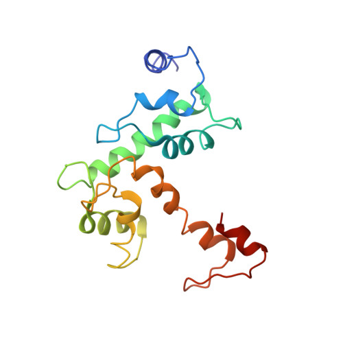
    ALG-2 (apoptosis-linked gene 2) is an apoptosis-linked calcium-binding protein with five EF-hand motifs in the C-terminal region. N-terminally truncated ALG-2 (des3-23ALG-2) was crystallized by the vapour-diffusion method in buffer consisting of either 50 mM MES pH 6.5, 12.5%(v/v) 2-propanol and 150 mM calcium acetate or 100 mM MES pH 6.0, 15%(v/v) ethanol and 200 mM zinc acetate. Crystals of the Ca(2+)-bound form belonged to space group P2(1)2(1)2(1), with unit-cell parameters a = 54.8, b = 154.4, c = 237.7 A, alpha = beta = gamma = 90 degrees , and diffracted to 3.1 A resolution. Crystals of the Zn(2+)-bound form belonged to space group P2(1)2(1)2(1), with unit-cell parameters a = 52.8, b = 147.5, c = 230.7 A, alpha = beta = gamma = 90 degrees , and diffracted to 3.3 A resolution. The structures of the Ca(2+)-bound form and the Zn(2+)-bound form were solved by the molecular-replacement method. Although both crystals contained eight ALG-2 molecules per asymmetric unit, the metal-ion locations and octameric arrangements were found to be significantly different.


  • Organizational Affiliation

    Department of Applied Molecular Biosciences, Graduate School of Bioagricultural Sciences, Nagoya University, Nagoya 464-8601, Japan.


Macromolecules
Find similar proteins by:  (by identity cutoff)  |  3D Structure
Entity ID: 1
MoleculeChains Sequence LengthOrganismDetailsImage
Programmed cell death protein 6
A, B, C, D, E
A, B, C, D, E, F, G, H
168Homo sapiensMutation(s): 0 
Gene Names: PDCD6ALG2
UniProt & NIH Common Fund Data Resources
Find proteins for O75340 (Homo sapiens)
Explore O75340 
Go to UniProtKB:  O75340
PHAROS:  O75340
GTEx:  ENSG00000249915 
Entity Groups  
Sequence Clusters30% Identity50% Identity70% Identity90% Identity95% Identity100% Identity
UniProt GroupO75340
Sequence Annotations
Expand
  • Reference Sequence
Small Molecules
Ligands 1 Unique
IDChains Name / Formula / InChI Key2D Diagram3D Interactions
CA
Query on CA

Download Ideal Coordinates CCD File 
AA [auth G]
BA [auth G]
CA [auth G]
DA [auth G]
EA [auth H]
AA [auth G],
BA [auth G],
CA [auth G],
DA [auth G],
EA [auth H],
FA [auth H],
GA [auth H],
I [auth A],
J [auth A],
K [auth B],
L [auth B],
M [auth B],
N [auth B],
O [auth C],
P [auth C],
Q [auth C],
R [auth D],
S [auth D],
T [auth D],
U [auth E],
V [auth E],
W [auth E],
X [auth F],
Y [auth F],
Z [auth F]
CALCIUM ION
Ca
BHPQYMZQTOCNFJ-UHFFFAOYSA-N
Experimental Data & Validation

Experimental Data

  • Method: X-RAY DIFFRACTION
  • Resolution: 3.10 Å
  • R-Value Free: 0.280 
  • R-Value Work: 0.222 
  • R-Value Observed: 0.228 
  • Space Group: P 21 21 21
Unit Cell:
Length ( Å )Angle ( ˚ )
a = 54.78α = 90
b = 154.425β = 90
c = 237.676γ = 90
Software Package:
Software NamePurpose
REFMACrefinement
HKL-2000data collection
HKL-2000data reduction
HKL-2000data scaling
MOLREPphasing

Structure Validation

View Full Validation Report


Entry History 

Deposition Data

Revision History  (Full details and data files)

  • Version 1.0: 2008-11-04
    Type: Initial release
  • Version 1.1: 2011-07-13
    Changes: Version format compliance
  • Version 1.2: 2017-10-11
    Changes: Refinement description
  • Version 1.3: 2023-11-01
    Changes: Data collection, Database references, Derived calculations, Refinement description