Crystal structure of Dioclea rostrata lectin: Insights into understanding the pH-dependent dimer-tetramer equilibrium and the structural basis for carbohydrate recognition in Diocleinae lectins
de Oliveira, T.M., Delatorre, P., da Rocha, B.A.M., de Souza, E.P., Nascimento, K.S., Bezerra, G.A., Moura, T.R., Benevides, R.G., Bezerra, E.H.S., Moreno, F.B.M.B., Freire, V.N., de Azevedo Jr., W.F., Cavada, B.S.(2008) J Struct Biol 164: 177-182
- PubMed: 18682294
- DOI: https://doi.org/10.1016/j.jsb.2008.05.012
- Primary Citation of Related Structures:
2ZBJ - PubMed Abstract:
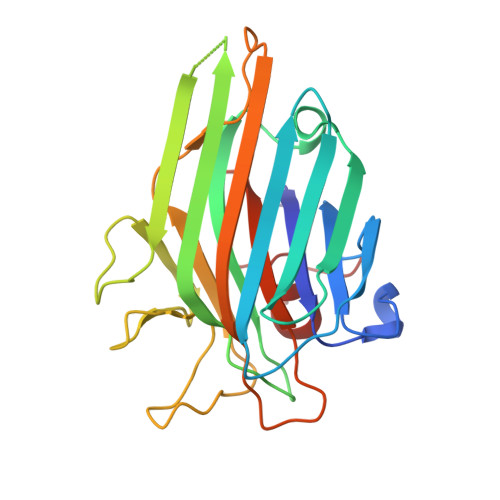
The legume lectins from the subtribe Diocleinae, often referred to as concanavalin A-like lectins, are a typical example of highly similar proteins that show distinct biological activities. The pH-dependent oligomerization that some of these lectins undergo and the relative position of amino acids within the carbohydrate-binding site are factors that have been reported to contribute to these differences in the activities of Diocleinae lectins. In the present work, we determined the amino acid sequence and the crystal structure of the lectin of Dioclea rostrata seeds (DRL), with the aim of investigating the structural bases of the different behavior displayed by this lectin in comparison to other Diocleinae lectins and determining the reason for the distinct pH-dependent dimer-tetramer equilibrium. In addition, we discovered a novel multimeric arrangement for this lectin.
Organizational Affiliation:
Federal University of Ceará, Departmento Biochemistry and Molecular Biology, Campus do Pici s/n Caixa Postal 6043, 60455-970 Fortaleza, Brazil.