


Crystal structure of cameline peptidoglycan recognition protein at 3.2 A resolution
Sharma, P., Jain, R., Singh, N., Sharma, S., Kaur, P., Singh, T.P.To be published.
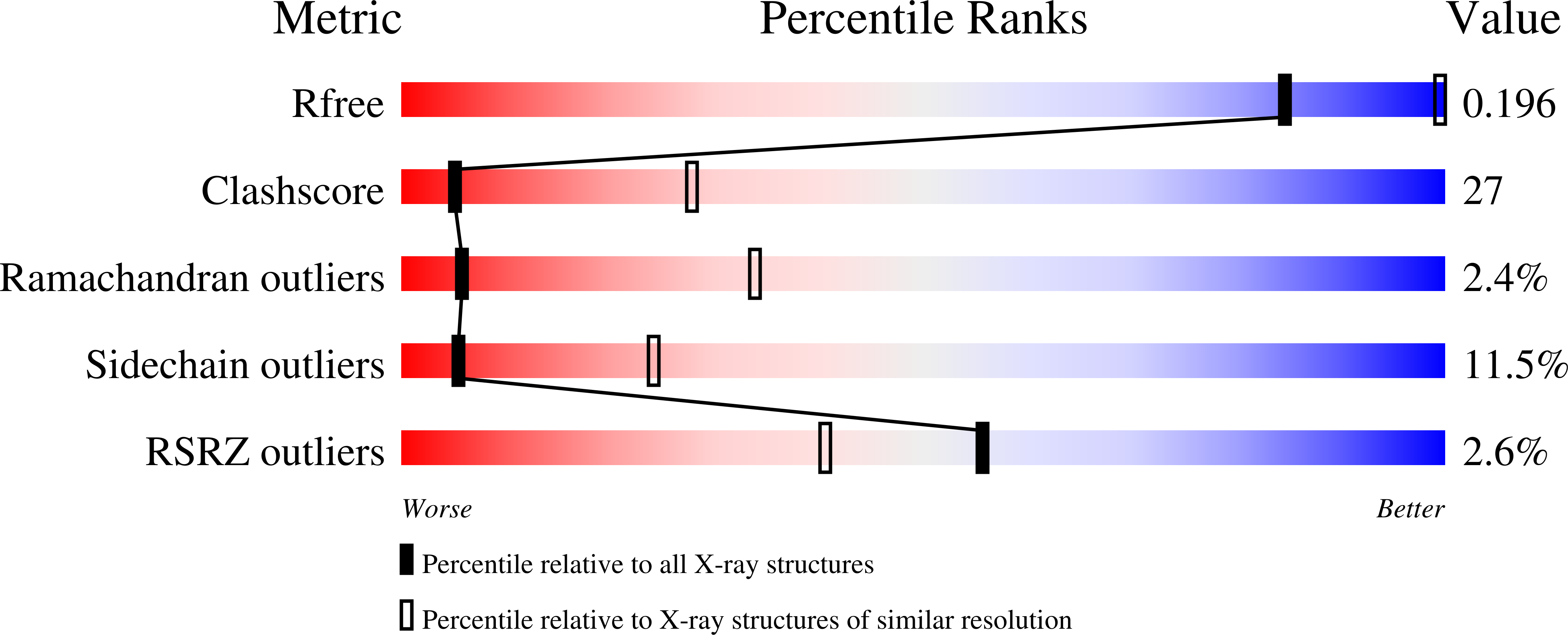
Experimental Data Snapshot
wwPDB Validation 3D Report Full Report
Entity ID: 1 | |||||
|---|---|---|---|---|---|
| Molecule | Chains | Sequence Length | Organism | Details | Image |
| Peptidoglycan recognition protein | 171 | Camelus dromedarius | Mutation(s): 0 | 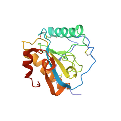 | |
UniProt | |||||
Find proteins for Q9GK12 (Camelus dromedarius) Explore Q9GK12 Go to UniProtKB: Q9GK12 | |||||
Entity Groups | |||||
| Sequence Clusters | 30% Identity50% Identity70% Identity90% Identity95% Identity100% Identity | ||||
| UniProt Group | Q9GK12 | ||||
Sequence AnnotationsExpand | |||||
| |||||
| Ligands 1 Unique | |||||
|---|---|---|---|---|---|
| ID | Chains | Name / Formula / InChI Key | 2D Diagram | 3D Interactions | |
| TLA Query on TLA | E [auth A], F [auth B], G [auth C] | L(+)-TARTARIC ACID C4 H6 O6 FEWJPZIEWOKRBE-JCYAYHJZSA-N |  | ||
| Length ( Å ) | Angle ( ˚ ) |
|---|---|
| a = 89.855 | α = 90 |
| b = 102.456 | β = 90 |
| c = 164.043 | γ = 90 |
| Software Name | Purpose |
|---|---|
| MAR345dtb | data collection |
| AMoRE | phasing |
| CNS | refinement |
| DENZO | data reduction |
| AUTOMAR | data reduction |
RCSB PDB (citation) is hosted by
RCSB PDB is a member of the

