2Z93

Crystal structure of Fab fragment of anti-ciguatoxin antibody 10C9 in complex with CTX3C-ABCD


Experimental Data Snapshot

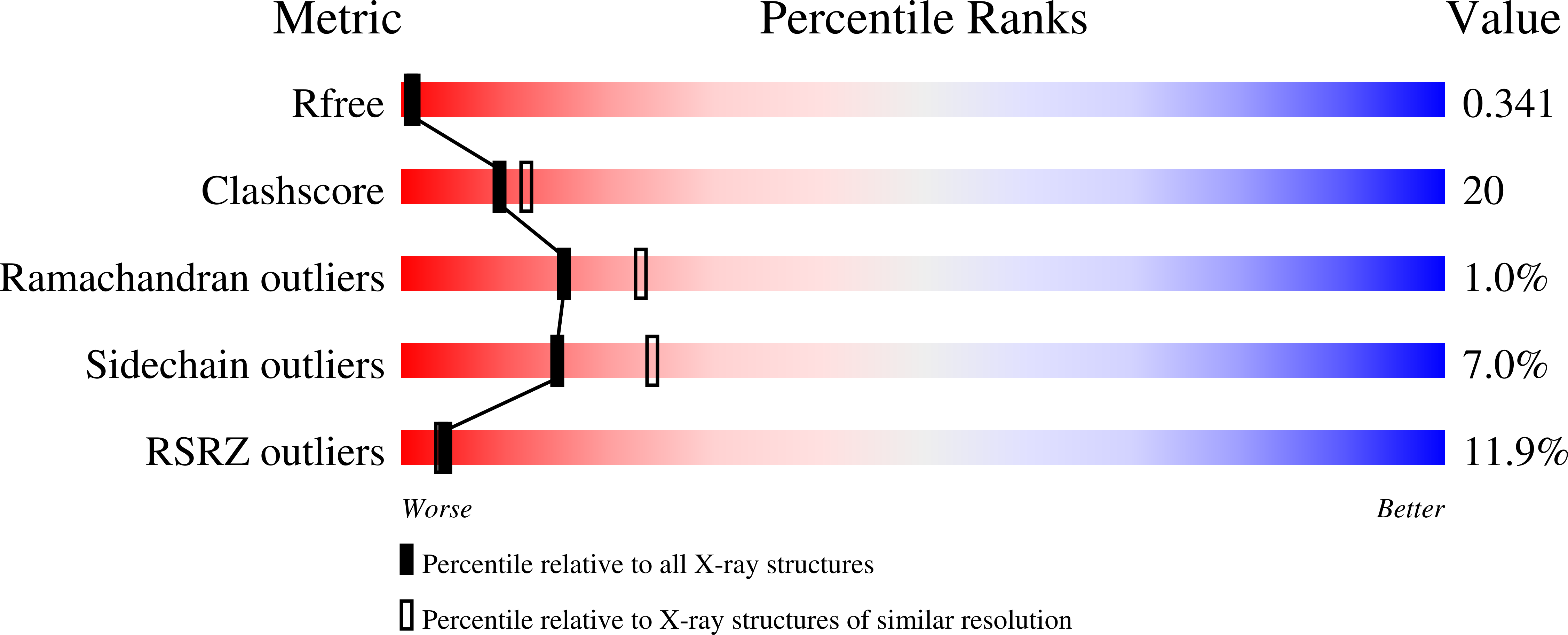
  • Method: X-RAY DIFFRACTION
  • Resolution: 2.40 Å
  • R-Value Free: 0.340 
  • R-Value Work: 0.297 

wwPDB Validation   3D Report Full Report


Ligand Structure Quality Assessment 


This is version 1.3 of the entry. See complete history


Literature

How Protein Recognizes Ladder-like Polycyclic Ethers: INTERACTIONS BETWEEN CIGUATOXIN (CTX3C) FRAGMENTS AND ITS SPECIFIC ANTIBODY 10C9

Ui, M.Tanaka, Y.Tsumuraya, T.Fujii, I.Inoue, M.Hirama, M.Tsumoto, K.

(2008) J Biol Chem 283: 19440-19447

  • DOI: https://doi.org/10.1074/jbc.M801282200
  • Primary Citation of Related Structures:  
    2Z91, 2Z92, 2Z93

  • PubMed Abstract: 

    Ciguatoxins are a family of marine toxins composed of transfused polycyclic ethers. It has not yet been clarified at the atomic level on the pathogenic mechanism of these toxins or the interaction between a polycyclic ether compounds and a protein. Using the crystal structures of anti-ciguatoxin antibody 10C9 Fab in ligand-free form and in complexes with ABCD-ring (CTX3C-ABCD) and ABCDE-ring (CTX3C-ABCDE) fragments of the antigen CTX3C at resolutions of 2.6, 2.4, and 2.3 angstroms, respectively, we elucidated the mechanism of the interaction between the polycyclic ethers and the antibody. 10C9 Fab has an extraordinarily large and deep binding pocket at the center of the variable region, where CTX3C-ABCD or CTX3C-ABCDE binds longitudinally in the pocket via hydrogen bonds and van der Waals interactions. Upon antigen-antibody complexation, 10C9 Fab adjusts to the antigen fragments by means of rotational motion in the variable region. In addition, the antigen fragment lacking the E-ring induces a large motion in the constant region. Consequently, the thermostability of 10C9 Fab is enhanced by 10 degrees C upon complexation with CTX3C-ABCDE but not with CTX3C-ABCD. The crystal structures presented in this study also show that 10C9 Fab recoginition of CTX3C antigens requires molecular rearrangements over the entire antibody structure. These results further expand the fundamental understanding of the mechanism by which ladder-like polycyclic ethers are recognized and may be useful for the design of novel therapeutic agents by antibodies, marine toxins, or new diagnostic reagents for the detection and targeting of members of the polycyclic ether family.


  • Organizational Affiliation

    Department of Medical Genome Sciences, Graduate School of Frontier Sciences, the University of Tokyo, Kashiwa 277-8562, Chiba, Japan.


Macromolecules
Find similar proteins by:  (by identity cutoff)  |  3D Structure
Entity ID: 1
MoleculeChains Sequence LengthOrganismDetailsImage
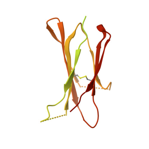
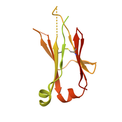
Anti-ciguatoxin antibody 10C9 Fab heavy chain
A, C
218Mus musculusMutation(s): 0 
Entity Groups  
Sequence Clusters30% Identity50% Identity70% Identity90% Identity95% Identity100% Identity
Sequence Annotations
Expand
  • Reference Sequence
Find similar proteins by:  (by identity cutoff)  |  3D Structure
Entity ID: 2
MoleculeChains Sequence LengthOrganismDetailsImage
Anti-ciguatoxin antibody 10C9 Fab light chain
B, D
213Mus musculusMutation(s): 0 
Entity Groups  
Sequence Clusters30% Identity50% Identity70% Identity90% Identity95% Identity100% Identity
Sequence Annotations
Expand
  • Reference Sequence
Small Molecules
Ligands 1 Unique
IDChains Name / Formula / InChI Key2D Diagram3D Interactions
END
Query on END

Download Ideal Coordinates CCD File 
E [auth C]1,6:5,9:8,12:11,16-TETRAANHYDRO-2,3,4,10,13,14-HEXADEOXY-D-GLYCERO-D-ALLO-D-GULO-HEPTADECA-2,13-DIENITOL
C17 H24 O7
VJZKPEAYONAPBB-MRBBLFKKSA-N
Binding Affinity Annotations 
IDSourceBinding Affinity
END Binding MOAD:  2Z93 Kd: 11.1 (nM) from 1 assay(s)
Experimental Data & Validation

Experimental Data

  • Method: X-RAY DIFFRACTION
  • Resolution: 2.40 Å
  • R-Value Free: 0.340 
  • R-Value Work: 0.297 
  • Space Group: P 1 21 1
Unit Cell:
Length ( Å )Angle ( ˚ )
a = 80.725α = 90
b = 45.41β = 98.9
c = 122.371γ = 90
Software Package:
Software NamePurpose
CNSrefinement
SERGUIdata collection
HKL-2000data reduction
HKL-2000data scaling
MOLREPphasing

Structure Validation

View Full Validation Report


Ligand Structure Quality Assessment 


Entry History 

Deposition Data

Revision History  (Full details and data files)

  • Version 1.0: 2008-05-06
    Type: Initial release
  • Version 1.1: 2011-07-13
    Changes: Version format compliance
  • Version 1.2: 2017-10-11
    Changes: Refinement description
  • Version 1.3: 2023-11-01
    Changes: Data collection, Database references, Derived calculations, Refinement description