


Crystal structure of PDZ domain from human Pleckstrin homology, Sec7
Kishishita, S., Nishino, A., Murayama, K., Terada, T., Shirouzu, M., Yokoyama, S.To be published.
Experimental Data Snapshot
wwPDB Validation 3D Report Full Report

Entity ID: 1 | |||||
|---|---|---|---|---|---|
| Molecule | Chains | Sequence Length | Organism | Details | Image |
| Pleckstrin homology Sec7 and coiled-coil domains-binding protein | 104 | Homo sapiens | Mutation(s): 0 | 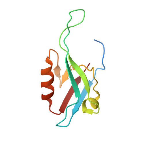 | |
UniProt & NIH Common Fund Data Resources | |||||
Find proteins for O60759 (Homo sapiens) Explore O60759 Go to UniProtKB: O60759 | |||||
PHAROS: O60759 GTEx: ENSG00000115165 | |||||
Entity Groups | |||||
| Sequence Clusters | 30% Identity50% Identity70% Identity90% Identity95% Identity100% Identity | ||||
| UniProt Group | O60759 | ||||
Sequence AnnotationsExpand | |||||
| |||||
| Length ( Å ) | Angle ( ˚ ) |
|---|---|
| a = 65.917 | α = 90 |
| b = 65.917 | β = 90 |
| c = 81.09 | γ = 120 |
| Software Name | Purpose |
|---|---|
| CNS | refinement |
| HKL-2000 | data reduction |
| HKL-2000 | data scaling |
| SOLVE | phasing |
RCSB PDB (citation) is hosted by
RCSB PDB is a member of the

