High-resolution Crystal Structure of {beta}2-Microglobulin Formed at pH 7.0
Iwata, K., Matsuura, T., Sakurai, K., Nakagawa, A., Goto, Y.(2007) J Biochem 142: 413-419
- PubMed: 17646174
- DOI: https://doi.org/10.1093/jb/mvm148
- Primary Citation of Related Structures:
2YXF - PubMed Abstract:
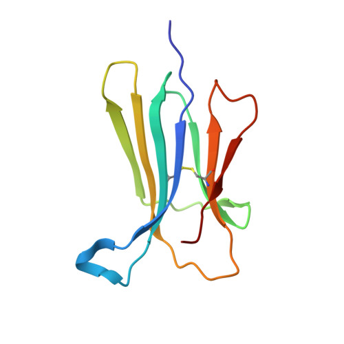
beta(2)-Microglobulin (beta2-m), a light chain of the major histocompatibility complex class I, forms amyloid fibrils in patients undergoing long-term haemodialysis, causing dialysis-related amyloidosis. Based on a comparison of the X-ray structure obtained at pH 5.7 and that of beta2-m in the histocompatibility complex, it has been proposed that the continuous D-strand observed in the crystal structure at pH 5.7 increases the propensity of beta2-m to self-associate via edge-to-edge interactions, thus initiating the formation of fibrils. To obtain further insight into the mechanism by which amyloid fibrils form, we determined the crystal structure of beta2-m at pH 7.0 at a resolution of up to 1.13 A. The crystal structure at pH 7.0 was basically the same as that at pH 5.6, suggesting that the conversion of the beta-bulge in strand D into a contiguous beta-strand is not unique to the crystals formed under slightly acidic conditions. In other words, although the formation of beta2-m fibrils was enhanced under acidic conditions, it remains unknown if it is related to the increased propensity for the disappearance of the beta-bulge in strand D. We consider that the enhanced fibrillation is more directly coupled with the decreased stability leading to the increased propensity of exposing amyloidogenic regions.
Organizational Affiliation:
Institute for Protein Research, Osaka University, and CREST, Japan Science and Technology Agency, Yamadaoka 3-2, Suita, Osaka, Japan.