2YNX

Crystal Structure of Ancestral Thioredoxin Relative to Last Archaea Common Ancestor (LACA) from the Precambrian Period


Experimental Data Snapshot

  • Method: X-RAY DIFFRACTION
  • Resolution: 1.75 Å
  • R-Value Free: 0.216 
  • R-Value Work: 0.160 
  • R-Value Observed: 0.163 

wwPDB Validation   3D Report Full Report


This is version 1.3 of the entry. See complete history


Literature

Conservation of Protein Structure Over Four Billion Years

Ingles-Prieto, A.Ibarra-Molero, B.Delgado-Delgado, A.Perez-Jimenez, R.Fernandez, J.M.Gaucher, E.A.Sanchez-Ruiz, J.M.Gavira, J.A.

(2013) Structure 21: 1690

  • DOI: https://doi.org/10.1016/j.str.2013.06.020
  • Primary Citation of Related Structures:  
    2YJ7, 2YN1, 2YNX, 2YOI, 2YPM, 3ZIV, 4BA7

  • PubMed Abstract: 

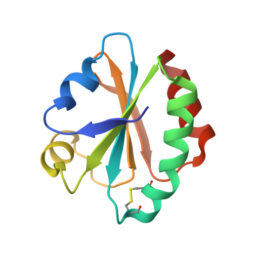
    Little is known about the evolution of protein structures and the degree of protein structure conservation over planetary time scales. Here, we report the X-ray crystal structures of seven laboratory resurrections of Precambrian thioredoxins dating up to approximately four billion years ago. Despite considerable sequence differences compared with extant enzymes, the ancestral proteins display the canonical thioredoxin fold, whereas only small structural changes have occurred over four billion years. This remarkable degree of structure conservation since a time near the last common ancestor of life supports a punctuated-equilibrium model of structure evolution in which the generation of new folds occurs over comparatively short periods and is followed by long periods of structural stasis.


  • Organizational Affiliation

    Departamento de Química Física, Facultad de Ciencias, Universidad de Granada, Granada 18071, Spain.


Macromolecules
Find similar proteins by:  (by identity cutoff)  |  3D Structure
Entity ID: 1
MoleculeChains Sequence LengthOrganismDetailsImage
LACA THIOREDOXIN
A, B
106synthetic constructMutation(s): 0 
EC: 1.8.1.9
Entity Groups  
Sequence Clusters30% Identity50% Identity70% Identity90% Identity95% Identity100% Identity
Sequence Annotations
Expand
  • Reference Sequence
Experimental Data & Validation

Experimental Data

  • Method: X-RAY DIFFRACTION
  • Resolution: 1.75 Å
  • R-Value Free: 0.216 
  • R-Value Work: 0.160 
  • R-Value Observed: 0.163 
  • Space Group: P 1
Unit Cell:
Length ( Å )Angle ( ˚ )
a = 32.153α = 90.76
b = 36.294β = 107.97
c = 48.118γ = 111.01
Software Package:
Software NamePurpose
PHENIXrefinement
XDSdata reduction
SCALAdata scaling
MOLREPphasing

Structure Validation

View Full Validation Report


Entry History 

Deposition Data

Revision History  (Full details and data files)

  • Version 1.0: 2013-08-21
    Type: Initial release
  • Version 1.1: 2013-09-25
    Changes: Database references
  • Version 1.2: 2019-03-06
    Changes: Data collection, Experimental preparation, Other
  • Version 1.3: 2023-12-20
    Changes: Data collection, Database references, Derived calculations, Other, Refinement description