2YKD

Structure of the matrix protein from human respiratory syncytial virus


Experimental Data Snapshot

  • Method: X-RAY DIFFRACTION
  • Resolution: 1.86 Å
  • R-Value Free: 0.229 
  • R-Value Work: 0.180 
  • R-Value Observed: 0.182 

wwPDB Validation   3D Report Full Report

Currently 2YKD does not have a validation slider image.


This is version 1.3 of the entry. See complete history


Literature

Influence of Lipids on the Interfacial Disposition of Respiratory Syncytical Virus Matrix Protein.

Mcphee, H.K.Carlisle, J.L.Beeby, A.Money, V.A.Watson, S.M.D.Yeo, R.P.Sanderson, J.M.

(2011) Langmuir 27: 304

  • DOI: https://doi.org/10.1021/la104041n
  • Primary Citation of Related Structures:  
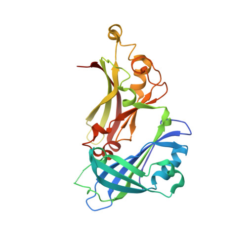
    2YKD

  • PubMed Abstract: 

    The propensity of a matrix protein from an enveloped virus of the Mononegavirales family to associate with lipids representative of the viral envelope has been determined using label-free methods, including tensiometry and Brewster angle microscopy on lipid films at the air-water interface and atomic force microscopy on monolayers transferred to OTS-treated silicon wafers. This has enabled factors that influence the disposition of the protein with respect to the lipid interface to be characterized. In the absence of sphingomyelin, respiratory syncytial virus matrix protein penetrates monolayers composed of mixtures of phosphocholines with phosphoethanolamines or cholesterol at the air-water interface. In ternary mixtures composed of sphingomyelin, 1,2-dioleoyl-sn-glycero-3-phosphocholine, and cholesterol, the protein exhibits two separate behaviors: (1) peripheral association with the surface of sphingomyelin-rich domains and (2) penetration of sphingomyelin-poor domains. Prolonged incubation of the protein with mixtures of phosphocholines and phosphoethanolamines leads to the formation of helical protein assemblies of uniform diameter that demonstrate an inherent propensity of the protein to assemble into a filamentous form.


  • Organizational Affiliation

    Department of Chemistry and Biophysical Sciences Institute, Durham University, South Road, Durham DH1 3LE, United Kingdom.


Macromolecules
Find similar proteins by:  (by identity cutoff)  |  3D Structure
Entity ID: 1
MoleculeChains Sequence LengthOrganismDetailsImage
MATRIX PROTEIN257human respiratory syncytial virusMutation(s): 0 
UniProt
Find proteins for P0DOE7 (Human respiratory syncytial virus A (strain A2))
Explore P0DOE7 
Go to UniProtKB:  P0DOE7
Entity Groups  
Sequence Clusters30% Identity50% Identity70% Identity90% Identity95% Identity100% Identity
UniProt GroupP0DOE7
Sequence Annotations
Expand
  • Reference Sequence
Experimental Data & Validation

Experimental Data

  • Method: X-RAY DIFFRACTION
  • Resolution: 1.86 Å
  • R-Value Free: 0.229 
  • R-Value Work: 0.180 
  • R-Value Observed: 0.182 
  • Space Group: C 1 2 1
Unit Cell:
Length ( Å )Angle ( ˚ )
a = 52.402α = 90
b = 78.902β = 96.18
c = 65.827γ = 90
Software Package:
Software NamePurpose
REFMACrefinement
MOSFLMdata reduction
SCALAdata scaling
AMoREphasing

Structure Validation

View Full Validation Report

Currently 2YKD does not have a validation slider image.


Entry History 

Deposition Data

Revision History  (Full details and data files)

  • Version 1.0: 2011-06-08
    Type: Initial release
  • Version 1.1: 2011-07-13
    Changes: Version format compliance
  • Version 1.2: 2019-05-08
    Changes: Data collection, Experimental preparation, Other
  • Version 1.3: 2023-12-20
    Changes: Data collection, Database references, Derived calculations, Other, Refinement description