Structural Basis for Electron and Methyl-Group Transfer in a Methyltransferase System Operating in the Reductive Acetyl-Coa Pathway
Goetzl, S., Jeoung, J.-H., Hennig, S.E., Dobbek, H.(2011) J Mol Biol 411: 96
- PubMed: 21640123
- DOI: https://doi.org/10.1016/j.jmb.2011.05.025
- Primary Citation of Related Structures:
2YCI, 2YCJ, 2YCK, 2YCL - PubMed Abstract:
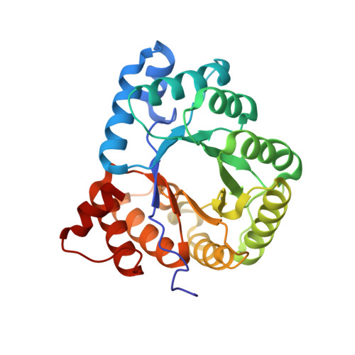
Several anaerobic acetogenic, methanogenic, hydrogenogenic, and sulfate-reducing microorganisms are able to use the reductive acetyl-CoA (Wood-Ljungdahl) pathway to convert CO₂ into biomass. The reductive acetyl-CoA pathway consists of two branches connected by the Co/Fe-containing corrinoid iron-sulfur protein (CoFeSP), which transfers a methyl group from a methyltransferase (MeTr)/methyltetrahydrofolate (CH₃-H₄ folate) complex to the reduced Ni-Ni-[4Fe-4S] cluster (cluster A) of acetyl-CoA synthase. We investigated the CoFeSP and MeTr couple of the hydrogenogenic bacterium Carboxydothermus hydrogenoformans and show that the two proteins are able to catalyze the methyl-group transfer reaction from CH₃-H₄ folate to the Co(I) center of CoFeSP. We determined the crystal structures of both proteins. The structure of CoFeSP includes the previously unresolved N-terminal domain of the large subunit of CoFeSP, revealing a unique four-helix-bundle-like architecture in which a [4Fe-4S] cluster is shielded by hydrophobic amino acids. It further reveals that the corrinoid and the [4Fe-4S] cluster binding domains are mobile, which is mandatory for the postulated electron transfer between them. Furthermore, we solved the crystal structures of apo-MeTr, CH₃-H₄-folate-bound MeTr, and H₄-folate-bound MeTr, revealing a substrate-induced closure of the CH₃-H₄ folate binding cavity of MeTr. We observed three different conformations of Asn200 depending on the substrate bound in the active site, demonstrating its conformational modulation by hydrogen-bonding interactions with the substrate. The observed flexibility could be essential to stabilize the transition state during methyl-group transfer. The conformational space and role of Asn200 are likely conserved in homologous cobalamin-dependent MeTrs such as methionine synthase.
Organizational Affiliation:
Institut für Biologie, Strukturbiologie/Biochemie, Humboldt-Universität zu Berlin, Leonor-Michaelis-Haus, 10115 Berlin, Germany.


