2XZS

Death associated protein kinase 1 residues 1-312


Experimental Data Snapshot

  • Method: X-RAY DIFFRACTION
  • Resolution: 2.00 Å
  • R-Value Free: 0.225 
  • R-Value Work: 0.183 
  • R-Value Observed: 0.185 

wwPDB Validation   3D Report Full Report


This is version 1.4 of the entry. See complete history


Literature

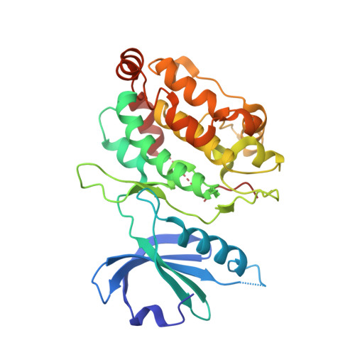
Death-Associated Protein Kinase Activity is Regulated by Coupled Calcium/Calmodulin Binding to Two Distinct Sites.

Simon, B.Huart, A.Temmerman, K.Vahokoski, J.Mertens, H.D.T.Komadina, D.Hoffmann, J.Yumerefendi, H.Svergun, D.I.Kursula, P.Schultz, C.Mccarthy, A.A.Hart, D.J.Wilmanns, M.

(2016) Structure 24: 851

  • DOI: https://doi.org/10.1016/j.str.2016.03.020
  • Primary Citation of Related Structures:  
    1YRP, 2A2A, 2XZS

  • PubMed Abstract: 

    The regulation of many protein kinases by binding to calcium/calmodulin connects two principal mechanisms in signaling processes: protein phosphorylation and responses to dose- and time-dependent calcium signals. We used the calcium/calmodulin-dependent members of the death-associated protein kinase (DAPK) family to investigate the role of a basic DAPK signature loop near the kinase active site. In DAPK2, this loop comprises a novel dimerization-regulated calcium/calmodulin-binding site, in addition to a well-established calcium/calmodulin site in the C-terminal autoregulatory domain. Unexpectedly, impairment of the basic loop interaction site completely abolishes calcium/calmodulin binding and DAPK2 activity is reduced to a residual level, indicative of coupled binding to the two sites. This contrasts with the generally accepted view that kinase calcium/calmodulin interactions are autonomous of the kinase catalytic domain. Our data establish an intricate model of multi-step kinase activation and expand our understanding of how calcium binding connects with other mechanisms involved in kinase activity regulation.


  • Organizational Affiliation

    European Molecular Biology Laboratory, Hamburg Unit, Notkestrasse 85, 22607 Hamburg, Germany.


Macromolecules
Find similar proteins by:  (by identity cutoff)  |  3D Structure
Entity ID: 1
MoleculeChains Sequence LengthOrganismDetailsImage
DEATH ASSOCIATED KINASE 1
A, B
312Homo sapiensMutation(s): 0 
EC: 2.7.11.1
UniProt & NIH Common Fund Data Resources
Find proteins for P53355 (Homo sapiens)
Explore P53355 
Go to UniProtKB:  P53355
PHAROS:  P53355
GTEx:  ENSG00000196730 
Entity Groups  
Sequence Clusters30% Identity50% Identity70% Identity90% Identity95% Identity100% Identity
UniProt GroupP53355
Sequence Annotations
Expand
  • Reference Sequence
Experimental Data & Validation

Experimental Data

  • Method: X-RAY DIFFRACTION
  • Resolution: 2.00 Å
  • R-Value Free: 0.225 
  • R-Value Work: 0.183 
  • R-Value Observed: 0.185 
  • Space Group: P 1 21 1
Unit Cell:
Length ( Å )Angle ( ˚ )
a = 56.3α = 90
b = 49.61β = 94.52
c = 130.5γ = 90
Software Package:
Software NamePurpose
REFMACrefinement
MOSFLMdata reduction
SCALAdata scaling
PHASERphasing

Structure Validation

View Full Validation Report


Entry History 

Deposition Data

Revision History  (Full details and data files)

  • Version 1.0: 2011-12-07
    Type: Initial release
  • Version 1.1: 2012-11-21
    Changes: Database references
  • Version 1.2: 2016-05-18
    Changes: Database references, Refinement description
  • Version 1.3: 2016-06-22
    Changes: Database references
  • Version 1.4: 2023-12-20
    Changes: Data collection, Database references, Derived calculations, Other, Refinement description