2X61

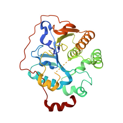
Crystal structure of the sialyltransferase CST-II in complex with trisaccharide acceptor and CMP


Experimental Data Snapshot

  • Method: X-RAY DIFFRACTION
  • Resolution: 1.95 Å
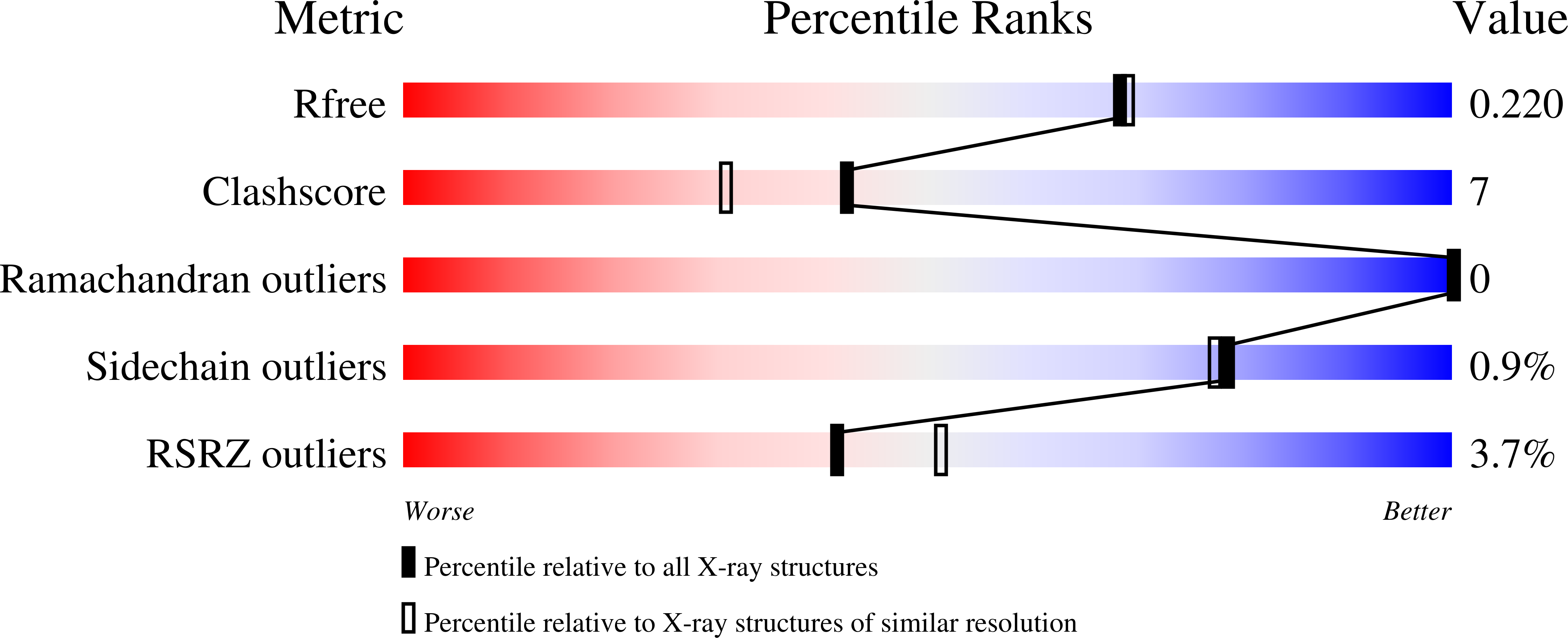
  • R-Value Free: 0.198 
  • R-Value Work: 0.164 
  • R-Value Observed: 0.166 

wwPDB Validation   3D Report Full Report


Ligand Structure Quality Assessment 


This is version 2.1 of the entry. See complete history


Literature

Structural and Kinetic Analysis of Substrate Binding to the Sialyltransferase Cst-II from Campylobacter Jejuni.

Lee, H.J.Lairson, L.L.Rich, J.R.Lameignere, E.Wakarchuk, W.W.Withers, S.G.Strynadka, N.C.

(2011) J Biol Chem 286: 35922

  • DOI: https://doi.org/10.1074/jbc.M111.261172
  • Primary Citation of Related Structures:  
    2X61, 2X62, 2X63

  • PubMed Abstract: 

    Sialic acids play important roles in various biological processes and typically terminate the oligosaccharide chains on the cell surfaces of a wide range of organisms, including mammals and bacteria. Their attachment is catalyzed by a set of sialyltransferases with defined specificities both for their acceptor sugars and the position of attachment. However, little is known of how this specificity is encoded. The structure of the bifunctional sialyltransferase Cst-II of the human pathogen Campylobacter jejuni in complex with CMP and the terminal trisaccharide of its natural acceptor (Neu5Ac-α-2,3-Gal-β-1,3-GalNAc) has been solved at 1.95 Å resolution, and its kinetic mechanism was shown to be iso-ordered Bi Bi, consistent with its dual acceptor substrate specificity. The trisaccharide acceptor is seen to bind to the active site of Cst-II through interactions primarily mediated by Asn-51, Tyr-81, and Arg-129. Kinetic and structural analyses of mutants modified at these positions indicate that these residues are critical for acceptor binding and catalysis, thereby providing significant new insight into the kinetic and catalytic mechanism, and acceptor specificity of this pathogen-encoded bifunctional GT-42 sialyltransferase.


  • Organizational Affiliation

    Department of Biochemistry and Molecular Biology, University of British Columbia, Vancouver, British Columbia V6T 1Z3; Centre for Blood Research, University of British Columbia, Vancouver, British Columbia V6T 1Z3.


Macromolecules
Find similar proteins by:  (by identity cutoff)  |  3D Structure
Entity ID: 1
MoleculeChains Sequence LengthOrganismDetailsImage
ALPHA-2,3-/2,8-SIALYLTRANSFERASE
A, B
258Campylobacter jejuniMutation(s): 2 
EC: 2.4.99
UniProt
Find proteins for Q9LAK3 (Campylobacter jejuni)
Explore Q9LAK3 
Go to UniProtKB:  Q9LAK3
Entity Groups  
Sequence Clusters30% Identity50% Identity70% Identity90% Identity95% Identity100% Identity
UniProt GroupQ9LAK3
Sequence Annotations
Expand
  • Reference Sequence
Oligosaccharides

Help

Entity ID: 2
MoleculeChains Length2D Diagram Glycosylation3D Interactions
N-acetyl-alpha-neuraminic acid-(2-3)-beta-D-galactopyranose-(1-3)-2-acetamido-2-deoxy-beta-D-galactopyranose
C, D
3N/A
Glycosylation Resources
GlyTouCan:  G02684WR
GlyCosmos:  G02684WR
GlyGen:  G02684WR
Small Molecules
Ligands 4 Unique
IDChains Name / Formula / InChI Key2D Diagram3D Interactions
CH
Query on CH

Download Ideal Coordinates CCD File 
G [auth A],
J [auth B]
N3-PROTONATED CYTIDINE-5'-MONOPHOSPHATE
C9 H15 N3 O8 P
ULTJSGLMABORQB-XVFCMESISA-O
MPD
Query on MPD

Download Ideal Coordinates CCD File 
F [auth A](4S)-2-METHYL-2,4-PENTANEDIOL
C6 H14 O2
SVTBMSDMJJWYQN-YFKPBYRVSA-N
EDO
Query on EDO

Download Ideal Coordinates CCD File 
I [auth A],
K [auth B]
1,2-ETHANEDIOL
C2 H6 O2
LYCAIKOWRPUZTN-UHFFFAOYSA-N
ACT
Query on ACT

Download Ideal Coordinates CCD File 
E [auth A],
H [auth A],
L [auth B]
ACETATE ION
C2 H3 O2
QTBSBXVTEAMEQO-UHFFFAOYSA-M
Experimental Data & Validation

Experimental Data

  • Method: X-RAY DIFFRACTION
  • Resolution: 1.95 Å
  • R-Value Free: 0.198 
  • R-Value Work: 0.164 
  • R-Value Observed: 0.166 
  • Space Group: P 4
Unit Cell:
Length ( Å )Angle ( ˚ )
a = 117.665α = 90
b = 117.665β = 90
c = 46.604γ = 90
Software Package:
Software NamePurpose
REFMACrefinement
HKL-2000data reduction
SCALEPACKdata scaling
PHASERphasing

Structure Validation

View Full Validation Report


Ligand Structure Quality Assessment 


Entry History 

Deposition Data

Revision History  (Full details and data files)

  • Version 1.0: 2011-03-02
    Type: Initial release
  • Version 1.1: 2011-08-17
    Changes: Atomic model, Derived calculations, Non-polymer description
  • Version 1.2: 2011-09-28
    Changes: Database references
  • Version 1.3: 2011-10-19
    Changes: Database references
  • Version 2.0: 2020-07-29
    Type: Remediation
    Reason: Carbohydrate remediation
    Changes: Advisory, Atomic model, Data collection, Derived calculations, Other, Structure summary
  • Version 2.1: 2023-12-20
    Changes: Data collection, Database references, Refinement description, Structure summary