Structure of the Papd-Papgii Pilin Complex Reveals an Open and Flexible P5 Pocket.
Ford, B.A., Verger, D., Dodson, K.W., Volkan, E., Kostakioti, M., Elam, J.S., Pinkner, J.S., Waksman, G., Hultgren, S.J.(2012) J Bacteriol 194: 6390
- PubMed: 23002225
- DOI: https://doi.org/10.1128/JB.06651-11
- Primary Citation of Related Structures:
2WMP - PubMed Abstract:
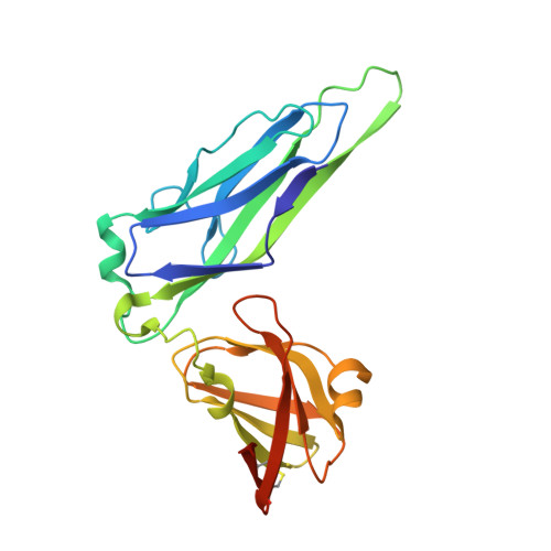
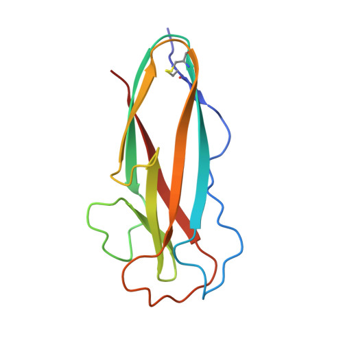
P pili are hairlike polymeric structures that mediate binding of uropathogenic Escherichia coli to the surface of the kidney via the PapG adhesin at their tips. PapG is composed of two domains: a lectin domain at the tip of the pilus followed by a pilin domain that comprises the initial polymerizing subunit of the 1,000-plus-subunit heteropolymeric pilus fiber. Prior to assembly, periplasmic pilin domains bind to a chaperone, PapD. PapD mediates donor strand complementation, in which a beta strand of PapD temporarily completes the pilin domain's fold, preventing premature, nonproductive interactions with other pilin subunits and facilitating subunit folding. Chaperone-subunit complexes are delivered to the outer membrane usher where donor strand exchange (DSE) replaces PapD's donated beta strand with an amino-terminal extension on the next incoming pilin subunit. This occurs via a zip-in-zip-out mechanism that initiates at a relatively accessible hydrophobic space termed the P5 pocket on the terminally incorporated pilus subunit. Here, we solve the structure of PapD in complex with the pilin domain of isoform II of PapG (PapGIIp). Our data revealed that PapGIIp adopts an immunoglobulin fold with a missing seventh strand, complemented in parallel by the G1 PapD strand, typical of pilin subunits. Comparisons with other chaperone-pilin complexes indicated that the interactive surfaces are highly conserved. Interestingly, the PapGIIp P5 pocket was in an open conformation, which, as molecular dynamics simulations revealed, switches between an open and a closed conformation due to the flexibility of the surrounding loops. Our study reveals the structural details of the DSE mechanism.
Organizational Affiliation:
Washington University Department of Pathology and Immunology, St. Louis, Missouri, USA.