2WHU

Fluorescent Protein mKeima at pH 8.0


Experimental Data Snapshot

  • Method: X-RAY DIFFRACTION
  • Resolution: 2.65 Å
  • R-Value Free: 0.307 
  • R-Value Work: 0.235 
  • R-Value Observed: 0.239 

wwPDB Validation   3D Report Full Report


This is version 2.1 of the entry. See complete history


Literature

Reverse Ph-Dependence of Chromophore Protonation Explains the Large Stokes Shift of the Red Fluorescent Protein Mkeima

Violot, S.Carpentier, P.Blanchoin, L.Bourgeois, D.

(2009) J Am Chem Soc 131: 10356

  • DOI: https://doi.org/10.1021/ja903695n
  • Primary Citation of Related Structures:  
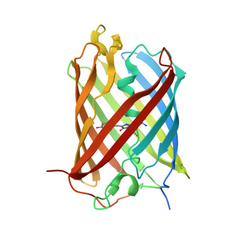
    2WHS, 2WHT, 2WHU

  • PubMed Abstract: 

    The recently developed red fluorescent protein Keima exhibits the largest Stokes shift (180 nm) observed to date. Combining X-ray crystallography with (in crystallo) UV-visible absorption, fluorescence, and Raman spectroscopy, we have investigated molecular determinants of this peculiar property. The results demonstrate a pH-dependent "reverse chromophore protonation" triggered by the key residue Asp157 and which couples to cis/trans isomerization of the chromophore. These data provided guidelines to rationally design a useful Keima variant.


  • Organizational Affiliation

    Institut de Recherches en Technologies et Sciences pour le Vivant, Laboratoire de Physiologie Cellulaire Végétale, CEA, CNRS, INRA, Université Joseph Fourier, 17 rue des Martyrs, F-38054 Grenoble, France.


Macromolecules
Find similar proteins by:  (by identity cutoff)  |  3D Structure
Entity ID: 1
MoleculeChains Sequence LengthOrganismDetailsImage
LARGE STOKES SHIFT FLUORESCENT PROTEIN
A, B, C, D
240Montipora sp. 20Mutation(s): 1 
UniProt
Find proteins for Q1JV70 (Montipora sp. 20)
Explore Q1JV70 
Go to UniProtKB:  Q1JV70
Entity Groups  
Sequence Clusters30% Identity50% Identity70% Identity90% Identity95% Identity100% Identity
UniProt GroupQ1JV70
Sequence Annotations
Expand
  • Reference Sequence
Small Molecules
Modified Residues  1 Unique
IDChains TypeFormula2D DiagramParent
CRQ
Query on CRQ
A, B, C, D
L-PEPTIDE LINKINGC16 H16 N4 O5GLN, TYR, GLY
Experimental Data & Validation

Experimental Data

  • Method: X-RAY DIFFRACTION
  • Resolution: 2.65 Å
  • R-Value Free: 0.307 
  • R-Value Work: 0.235 
  • R-Value Observed: 0.239 
  • Space Group: C 1 2 1
Unit Cell:
Length ( Å )Angle ( ˚ )
a = 225.11α = 90
b = 57.75β = 108.99
c = 93.07γ = 90
Software Package:
Software NamePurpose
REFMACrefinement
XDSdata reduction
XSCALEdata scaling
PHASERphasing

Structure Validation

View Full Validation Report


Entry History 

Deposition Data

Revision History  (Full details and data files)

  • Version 1.0: 2009-08-11
    Type: Initial release
  • Version 1.1: 2012-07-04
    Changes: Database references, Derived calculations, Non-polymer description, Other, Version format compliance
  • Version 1.2: 2018-03-07
    Changes: Source and taxonomy
  • Version 1.3: 2019-03-06
    Changes: Data collection, Derived calculations, Experimental preparation
  • Version 1.4: 2019-10-23
    Changes: Data collection, Database references, Other
  • Version 2.0: 2023-11-15
    Changes: Advisory, Atomic model, Data collection, Database references, Derived calculations
  • Version 2.1: 2023-12-13
    Changes: Refinement description