2WB3

The partial structure of a group A streptococcal phage-encoded tail fibre hyaluronate lyase Hylp3


Experimental Data Snapshot

  • Method: X-RAY DIFFRACTION
  • Resolution: 1.90 Å
  • R-Value Free: 0.242 
  • R-Value Work: 0.190 
  • R-Value Observed: 0.192 

wwPDB Validation   3D Report Full Report


This is version 1.2 of the entry. See complete history


Literature

Structures of Two Truncated Phage-Tail Hyaluronate Lyases from Streptococcus Pyogenes Serotype M1.

Martinez-Fleites, C.Smith, N.L.Turkenburg, J.P.Black, G.W.Taylor, E.J.

(2009) Acta Crystallogr Sect F Struct Biol Cryst Commun 65: 963

  • DOI: https://doi.org/10.1107/S1744309109032813
  • Primary Citation of Related Structures:  
    2WB3, 2WH7

  • PubMed Abstract: 

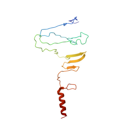
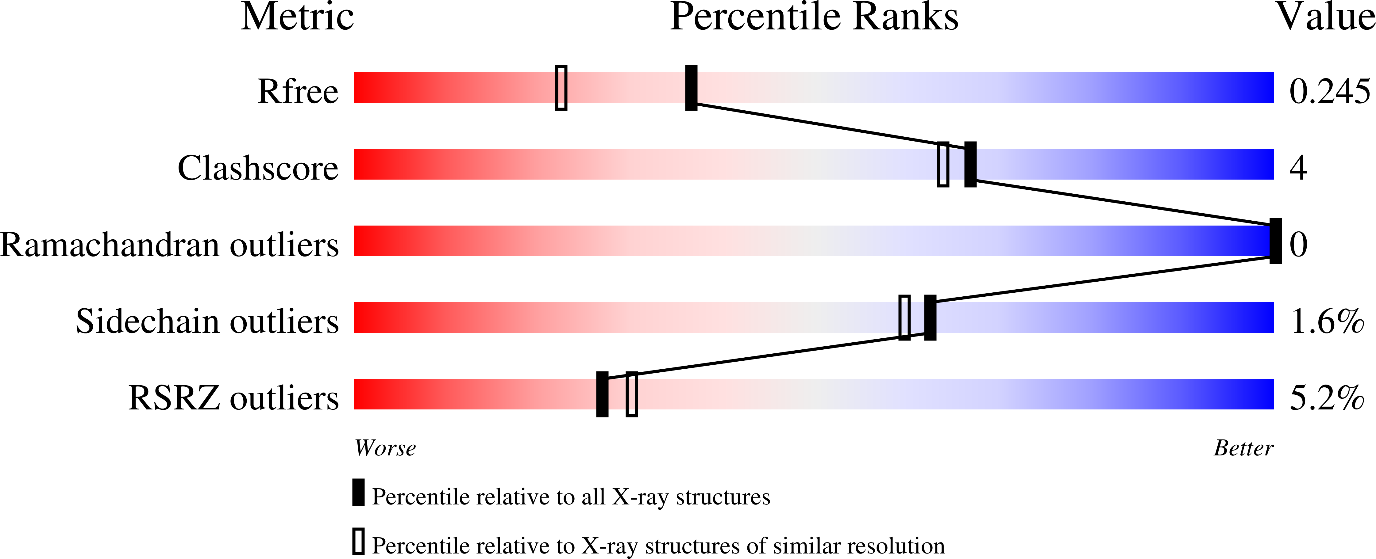
    The crystal structures of truncated forms of the Streptococcus pyogenes phage-encoded hyaluronate lyases HylP2 and HylP3 were determined by molecular replacement to 1.6 and 1.9 A resolution, respectively. The truncated forms crystallized in a hexagonal space group, forming a trimer around the threefold crystallographic axis. The arrangement of the fold is very similar to that observed in the structure of the related hyaluronate lyase HylP1. The structural elements putatively involved in substrate recognition are found to be conserved in both the HylP2 and HylP3 fragments.


  • Organizational Affiliation

    Structural Biology Laboratory, Department of Chemistry, The University of York, York YO10 5YW, England.


Macromolecules
Find similar proteins by:  (by identity cutoff)  |  3D Structure
Entity ID: 1
MoleculeChains Sequence LengthOrganismDetailsImage
HYALURONIDASE-PHAGE ASSOCIATED157Streptococcus pyogenesMutation(s): 0 
EC: 4.2.2.1
UniProt
Find proteins for Q99Z19 (Streptococcus pyogenes serotype M1)
Explore Q99Z19 
Go to UniProtKB:  Q99Z19
Entity Groups  
Sequence Clusters30% Identity50% Identity70% Identity90% Identity95% Identity100% Identity
UniProt GroupQ99Z19
Sequence Annotations
Expand
  • Reference Sequence
Experimental Data & Validation

Experimental Data

  • Method: X-RAY DIFFRACTION
  • Resolution: 1.90 Å
  • R-Value Free: 0.242 
  • R-Value Work: 0.190 
  • R-Value Observed: 0.192 
  • Space Group: P 63 2 2
Unit Cell:
Length ( Å )Angle ( ˚ )
a = 49.476α = 90
b = 49.476β = 90
c = 241.247γ = 120
Software Package:
Software NamePurpose
REFMACrefinement
DENZOdata reduction
SCALEPACKdata scaling
PHASERphasing

Structure Validation

View Full Validation Report


Entry History 

Deposition Data

Revision History  (Full details and data files)

  • Version 1.0: 2009-09-01
    Type: Initial release
  • Version 1.1: 2011-05-08
    Changes: Version format compliance
  • Version 1.2: 2011-07-13
    Changes: Version format compliance