2W9E

Structure of ICSM 18 (anti-Prp therapeutic antibody) Fab fragment complexed with human Prp fragment 119-231


Experimental Data Snapshot

  • Method: X-RAY DIFFRACTION
  • Resolution: 2.90 Å
  • R-Value Free: 0.269 
  • R-Value Work: 0.207 
  • R-Value Observed: 0.210 

wwPDB Validation   3D Report Full Report


This is version 1.2 of the entry. See complete history


Literature

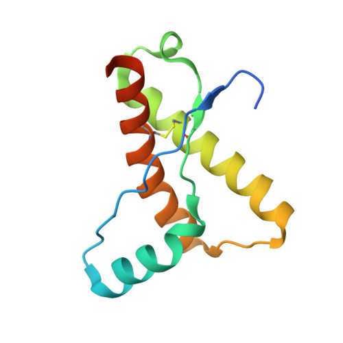
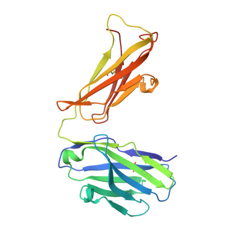
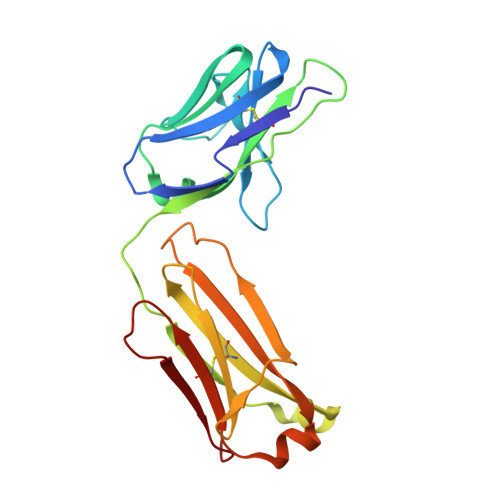
Crystal Structure of Human Prion Protein Bound to a Therapeutic Antibody.

Antonyuk, S.V.Trevitt, C.R.Strange, R.W.Jackson, G.S.Sangar, D.Batchelor, M.Cooper, S.Fraser, C.Jones, S.Georgiou, T.Khalili-Shirazi, A.Clarke, A.R.Hasnain, S.S.Collinge, J.

(2009) Proc Natl Acad Sci U S A 106: 2554

  • DOI: https://doi.org/10.1073/pnas.0809170106
  • Primary Citation of Related Structures:  
    2W9D, 2W9E

  • PubMed Abstract: 

    Prion infection is characterized by the conversion of host cellular prion protein (PrP(C)) into disease-related conformers (PrP(Sc)) and can be arrested in vivo by passive immunization with anti-PrP monoclonal antibodies. Here, we show that the ability of an antibody to cure prion-infected cells correlates with its binding affinity for PrP(C) rather than PrP(Sc). We have visualized this interaction at the molecular level by determining the crystal structure of human PrP bound to the Fab fragment of monoclonal antibody ICSM 18, which has the highest affinity for PrP(C) and the highest therapeutic potency in vitro and in vivo. In this crystal structure, human PrP is observed in its native PrP(C) conformation. Interactions between neighboring PrP molecules in the crystal structure are mediated by close homotypic contacts between residues at position 129 that lead to the formation of a 4-strand intermolecular beta-sheet. The importance of this residue in mediating protein-protein contact could explain the genetic susceptibility and prion strain selection determined by polymorphic residue 129 in human prion disease, one of the strongest common susceptibility polymorphisms known in any human disease.


  • Organizational Affiliation

    Molecular Biophysics Group, Science and Technology Facilities Council, Daresbury Laboratory, Warrington, Cheshire WA4 4AD, United Kingdom.


Macromolecules
Find similar proteins by:  (by identity cutoff)  |  3D Structure
Entity ID: 1
MoleculeChains Sequence LengthOrganismDetailsImage
MAJOR PRION PROTEIN113Homo sapiensMutation(s): 0 
UniProt & NIH Common Fund Data Resources
Find proteins for P04156 (Homo sapiens)
Explore P04156 
Go to UniProtKB:  P04156
PHAROS:  P04156
GTEx:  ENSG00000171867 
Entity Groups  
Sequence Clusters30% Identity50% Identity70% Identity90% Identity95% Identity100% Identity
UniProt GroupP04156
Sequence Annotations
Expand
  • Reference Sequence
Find similar proteins by:  (by identity cutoff)  |  3D Structure
Entity ID: 2
MoleculeChains Sequence LengthOrganismDetailsImage
ICSM 18-ANTI-PRP THERAPEUTIC FAB HEAVY CHAINB [auth H]215Mus musculusMutation(s): 0 
Entity Groups  
Sequence Clusters30% Identity50% Identity70% Identity90% Identity95% Identity100% Identity
Sequence Annotations
Expand
  • Reference Sequence
Find similar proteins by:  (by identity cutoff)  |  3D Structure
Entity ID: 3
MoleculeChains Sequence LengthOrganismDetailsImage
ICSM 18-ANTI-PRP THERAPEUTIC FAB LIGHT CHAINC [auth L]212Mus musculusMutation(s): 0 
Entity Groups  
Sequence Clusters30% Identity50% Identity70% Identity90% Identity95% Identity100% Identity
Sequence Annotations
Expand
  • Reference Sequence
Small Molecules
Ligands 1 Unique
IDChains Name / Formula / InChI Key2D Diagram3D Interactions
SO4
Query on SO4

Download Ideal Coordinates CCD File 
D [auth A]SULFATE ION
O4 S
QAOWNCQODCNURD-UHFFFAOYSA-L
Experimental Data & Validation

Experimental Data

  • Method: X-RAY DIFFRACTION
  • Resolution: 2.90 Å
  • R-Value Free: 0.269 
  • R-Value Work: 0.207 
  • R-Value Observed: 0.210 
  • Space Group: P 63 2 2
Unit Cell:
Length ( Å )Angle ( ˚ )
a = 126.149α = 90
b = 126.149β = 90
c = 134.126γ = 120
Software Package:
Software NamePurpose
REFMACrefinement
HKL-2000data reduction
HKL-2000data scaling
PHASERphasing

Structure Validation

View Full Validation Report


Entry History 

Revision History  (Full details and data files)

  • Version 1.0: 2009-02-03
    Type: Initial release
  • Version 1.1: 2011-07-13
    Changes: Advisory, Refinement description, Version format compliance
  • Version 1.2: 2023-12-13
    Changes: Data collection, Database references, Derived calculations, Other, Refinement description