2W99

Crystal Structure of CDK4 in complex with a D-type cyclin


Experimental Data Snapshot

  • Method: X-RAY DIFFRACTION
  • Resolution: 2.80 Å
  • R-Value Free: 0.270 
  • R-Value Work: 0.213 
  • R-Value Observed: 0.216 

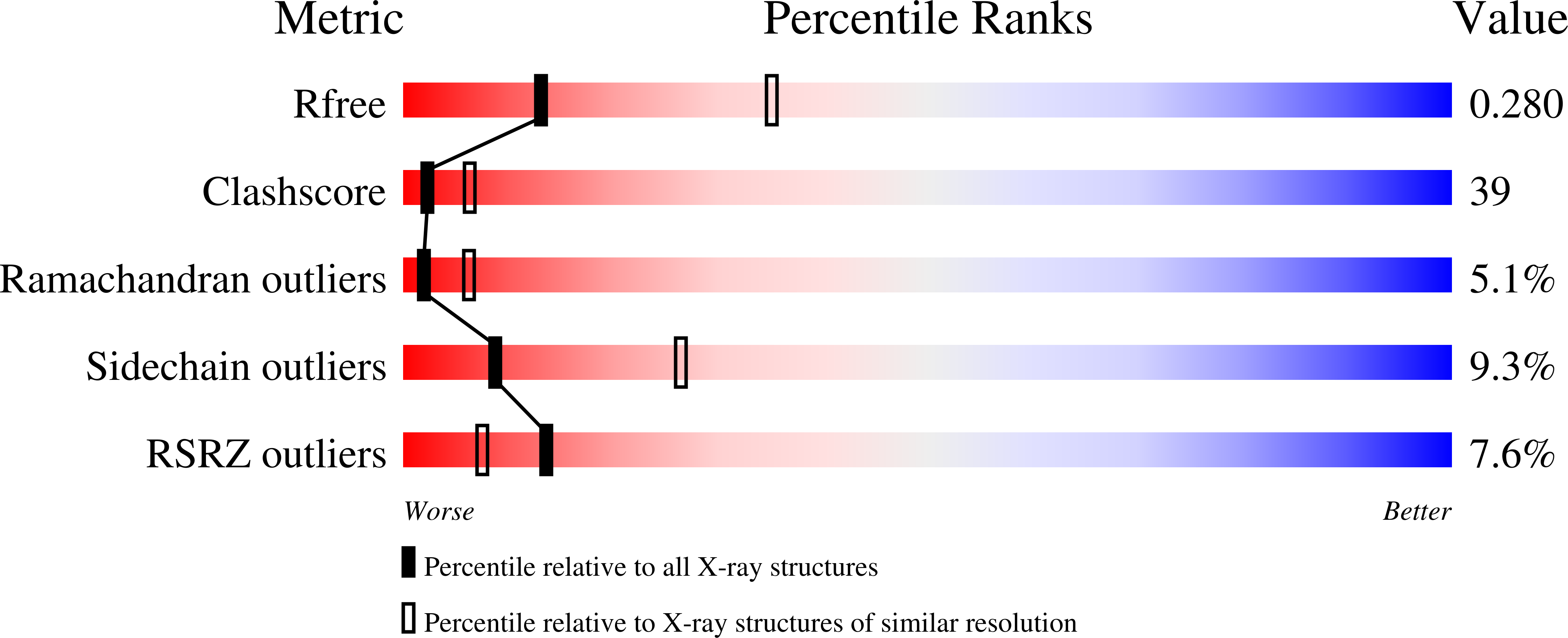
wwPDB Validation   3D Report Full Report


This is version 1.4 of the entry. See complete history


Literature

Crystal Structure of Human Cdk4 in Complex with a D-Type Cyclin.

Day, P.J.Cleasby, A.Tickle, I.J.O'Reilly, M.Coyle, J.E.Holding, F.P.Mcmenamin, R.L.Yon, J.Chopra, R.Lengauer, C.Jhoti, H.

(2009) Proc Natl Acad Sci U S A 106: 4166

  • DOI: https://doi.org/10.1073/pnas.0809645106
  • Primary Citation of Related Structures:  
    2W96, 2W99, 2W9F, 2W9Z

  • PubMed Abstract: 

    The cyclin D1-cyclin-dependent kinase 4 (CDK4) complex is a key regulator of the transition through the G(1) phase of the cell cycle. Among the cyclin/CDKs, CDK4 and cyclin D1 are the most frequently activated by somatic genetic alterations in multiple tumor types. Thus, aberrant regulation of the CDK4/cyclin D1 pathway plays an essential role in oncogenesis; hence, CDK4 is a genetically validated therapeutic target. Although X-ray crystallographic structures have been determined for various CDK/cyclin complexes, CDK4/cyclin D1 has remained highly refractory to structure determination. Here, we report the crystal structure of CDK4 in complex with cyclin D1 at a resolution of 2.3 A. Although CDK4 is bound to cyclin D1 and has a phosphorylated T-loop, CDK4 is in an inactive conformation and the conformation of the heterodimer diverges from the previously known CDK/cyclin binary complexes, which suggests a unique mechanism for the process of CDK4 regulation and activation.


  • Organizational Affiliation

    Astex Therapeutics Ltd., 436 Cambridge Science Park, Milton Road, Cambridge CB4 0QA, United Kingdom.


Macromolecules
Find similar proteins by:  (by identity cutoff)  |  3D Structure
Entity ID: 1
MoleculeChains Sequence LengthOrganismDetailsImage
G1/S-SPECIFIC CYCLIN-D1271Homo sapiensMutation(s): 0 
UniProt & NIH Common Fund Data Resources
Find proteins for P24385 (Homo sapiens)
Explore P24385 
Go to UniProtKB:  P24385
PHAROS:  P24385
GTEx:  ENSG00000110092 
Entity Groups  
Sequence Clusters30% Identity50% Identity70% Identity90% Identity95% Identity100% Identity
UniProt GroupP24385
Sequence Annotations
Expand
  • Reference Sequence
Find similar proteins by:  (by identity cutoff)  |  3D Structure
Entity ID: 2
MoleculeChains Sequence LengthOrganismDetailsImage
CELL DIVISION PROTEIN KINASE 4306Homo sapiensMutation(s): 3 
EC: 2.7.11.22
UniProt & NIH Common Fund Data Resources
Find proteins for P11802 (Homo sapiens)
Explore P11802 
Go to UniProtKB:  P11802
PHAROS:  P11802
GTEx:  ENSG00000135446 
Entity Groups  
Sequence Clusters30% Identity50% Identity70% Identity90% Identity95% Identity100% Identity
UniProt GroupP11802
Sequence Annotations
Expand
  • Reference Sequence
Experimental Data & Validation

Experimental Data

  • Method: X-RAY DIFFRACTION
  • Resolution: 2.80 Å
  • R-Value Free: 0.270 
  • R-Value Work: 0.213 
  • R-Value Observed: 0.216 
  • Space Group: P 21 21 21
Unit Cell:
Length ( Å )Angle ( ˚ )
a = 57.023α = 90
b = 64.684β = 90
c = 188.747γ = 90
Software Package:
Software NamePurpose
BUSTER-TNTrefinement
MOSFLMdata reduction
SCALAdata scaling
PHASERphasing

Structure Validation

View Full Validation Report


Entry History 

Deposition Data

Revision History  (Full details and data files)

  • Version 1.0: 2009-03-10
    Type: Initial release
  • Version 1.1: 2011-05-08
    Changes: Version format compliance
  • Version 1.2: 2011-07-13
    Changes: Version format compliance
  • Version 1.3: 2019-04-24
    Changes: Data collection, Other, Source and taxonomy
  • Version 1.4: 2023-12-13
    Changes: Data collection, Database references, Other, Refinement description