2W89

Crystal structure of the E.coli tRNAArg aminoacyl stem issoacceptor RR-1660 at 2.0 Angstroem resolution


Experimental Data Snapshot

  • Method: X-RAY DIFFRACTION
  • Resolution: 2.00 Å
  • R-Value Free: 0.255 
  • R-Value Work: 0.212 
  • R-Value Observed: 0.214 

wwPDB Validation   3D Report Full Report


This is version 1.3 of the entry. See complete history


Literature

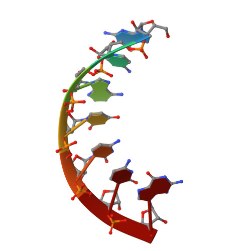
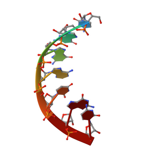
Crystal Structure of the E. Coli tRNA(Arg) Aminoacyl Stem Isoacceptor Rr-1660 at 2.0 A Resolution.

Eichert, A.Perbandt, M.Oberthur, D.Schreiber, A.Furste, J.P.Betzel, C.Erdmann, V.A.Forster, C.

(2009) Biochem Biophys Res Commun 385: 84

  • DOI: https://doi.org/10.1016/j.bbrc.2009.04.158
  • Primary Citation of Related Structures:  
    2W89

  • PubMed Abstract: 

    Due to the redundancy of the genetic code there exist six mRNA codons for arginine and several tRNA(Arg) isoacceptors which translate these triplets to protein within the context of the mRNA. The tRNA identity elements assure the correct aminoacylation of the tRNA with the cognate amino acid by the aminoacyl-tRNA-synthetases. In tRNA(Arg), the identity elements consist of the anticodon, parts of the D-loop and the discriminator base. The minor groove of the acceptor stem interacts with the arginyl-tRNA-synthetase. We crystallized different Escherichia coli tRNA(Arg) acceptor stem helices and solved the structure of the tRNA(Arg) isoacceptor RR-1660 microhelix by X-ray structure analysis. The acceptor stem helix crystallizes in the space group P1 with the cell constants a=26.28, b=28.92, c=29.00 A, alpha=105.74, beta=99.01, gamma=97.44 degrees and two molecules per asymmetric unit. The RNA hydration pattern consists of 88 bound water molecules. Additionally, one glycerol molecule is bound within the interface of the two RNA molecules.


  • Organizational Affiliation

    Institute of Chemistry and Biochemistry, Free University Berlin, Thielallee 63, 14195 Berlin, Germany.


Macromolecules

Find similar nucleic acids by:  Sequence   |   3D Structure  

Entity ID: 1
MoleculeChains LengthOrganismImage
5'-R(*GP*CP*AP*UP*CP*CP*GP)-3'
A, C
7Escherichia coli
Sequence Annotations
Expand
  • Reference Sequence

Find similar nucleic acids by:  Sequence   |   3D Structure  

Entity ID: 2
MoleculeChains LengthOrganismImage
5'-R(*CP*GP*GP*AP*UP*GP*CP)-3'
B, D
7Escherichia coli
Sequence Annotations
Expand
  • Reference Sequence
Small Molecules
Ligands 1 Unique
IDChains Name / Formula / InChI Key2D Diagram3D Interactions
GOL
Query on GOL

Download Ideal Coordinates CCD File 
E [auth B]GLYCEROL
C3 H8 O3
PEDCQBHIVMGVHV-UHFFFAOYSA-N
Experimental Data & Validation

Experimental Data

  • Method: X-RAY DIFFRACTION
  • Resolution: 2.00 Å
  • R-Value Free: 0.255 
  • R-Value Work: 0.212 
  • R-Value Observed: 0.214 
  • Space Group: P 1
Unit Cell:
Length ( Å )Angle ( ˚ )
a = 26.368α = 105.89
b = 28.984β = 98.76
c = 29.134γ = 97.85
Software Package:
Software NamePurpose
REFMACrefinement
DENZOdata reduction
SCALEPACKdata scaling
PHASERphasing

Structure Validation

View Full Validation Report


Entry History 

Deposition Data

Revision History  (Full details and data files)

  • Version 1.0: 2009-11-17
    Type: Initial release
  • Version 1.1: 2015-01-28
    Changes: Derived calculations, Non-polymer description, Refinement description, Version format compliance
  • Version 1.2: 2019-05-08
    Changes: Data collection, Experimental preparation, Other
  • Version 1.3: 2023-12-13
    Changes: Data collection, Database references, Derived calculations, Other, Refinement description