2W1E

Structure determination of Aurora Kinase in complex with inhibitor


Experimental Data Snapshot

  • Method: X-RAY DIFFRACTION
  • Resolution: 2.93 Å
  • R-Value Free: 0.302 
  • R-Value Work: 0.233 
  • R-Value Observed: 0.237 

wwPDB Validation   3D Report Full Report


Ligand Structure Quality Assessment 


This is version 1.3 of the entry. See complete history


Literature

Fragment-Based Discovery of the Pyrazol-4-Yl Urea (at9283), a Multitargeted Kinase Inhibitor with Potent Aurora Kinase Activity.

Howard, S.Berdini, V.Boulstridge, J.A.Carr, M.G.Cross, D.M.Curry, J.Devine, L.A.Early, T.R.Fazal, L.Gill, A.L.Heathcote, M.Maman, S.Matthews, J.E.Mcmenamin, R.L.Navarro, E.F.O'Brien, M.A.O'Reilly, M.Rees, D.C.Reule, M.Tisi, D.Williams, G.Vinkovic, M.Wyatt, P.G.

(2009) J Med Chem 52: 379

  • DOI: https://doi.org/10.1021/jm800984v
  • Primary Citation of Related Structures:  
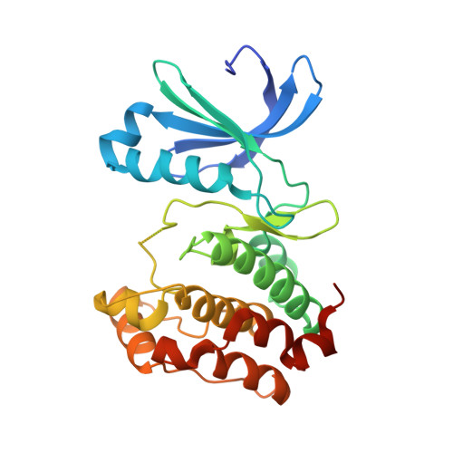
    2W1C, 2W1D, 2W1E, 2W1F, 2W1G, 2W1H, 2W1I

  • PubMed Abstract: 

    Here, we describe the identification of a clinical candidate via structure-based optimization of a ligand efficient pyrazole-benzimidazole fragment. Aurora kinases play a key role in the regulation of mitosis and in recent years have become attractive targets for the treatment of cancer. X-ray crystallographic structures were generated using a novel soakable form of Aurora A and were used to drive the optimization toward potent (IC(50) approximately 3 nM) dual Aurora A/Aurora B inhibitors. These compounds inhibited growth and survival of HCT116 cells and produced the polyploid cellular phenotype typically associated with Aurora B kinase inhibition. Optimization of cellular activity and physicochemical properties ultimately led to the identification of compound 16 (AT9283). In addition to Aurora A and Aurora B, compound 16 was also found to inhibit a number of other kinases including JAK2 and Abl (T315I). This compound demonstrated in vivo efficacy in mouse xenograft models and is currently under evaluation in phase I clinical trials.


  • Organizational Affiliation

    Astex Therapeutics Ltd., 436 Cambridge Science Park, Milton Road, Cambridge, CB4 0QA, UK. s.howard@astex-therapeutics.com


Macromolecules
Find similar proteins by:  (by identity cutoff)  |  3D Structure
Entity ID: 1
MoleculeChains Sequence LengthOrganismDetailsImage
SERINE/THREONINE-PROTEIN KINASE 6275Homo sapiensMutation(s): 0 
EC: 2.7.11.1
UniProt & NIH Common Fund Data Resources
Find proteins for O14965 (Homo sapiens)
Explore O14965 
Go to UniProtKB:  O14965
PHAROS:  O14965
GTEx:  ENSG00000087586 
Entity Groups  
Sequence Clusters30% Identity50% Identity70% Identity90% Identity95% Identity100% Identity
UniProt GroupO14965
Sequence Annotations
Expand
  • Reference Sequence
Small Molecules
Ligands 1 Unique
IDChains Name / Formula / InChI Key2D Diagram3D Interactions
L0E
Query on L0E

Download Ideal Coordinates CCD File 
B [auth A]4-[(2-{4-[(PHENYLCARBAMOYL)AMINO]-1H-PYRAZOL-3-YL}-1H-BENZIMIDAZOL-5-YL)METHYL]MORPHOLIN-4-IUM
C22 H24 N7 O2
SOTHNRIRXPUQEZ-UHFFFAOYSA-O
Binding Affinity Annotations 
IDSourceBinding Affinity
L0E PDBBind:  2W1E IC50: 12 (nM) from 1 assay(s)
Experimental Data & Validation

Experimental Data

  • Method: X-RAY DIFFRACTION
  • Resolution: 2.93 Å
  • R-Value Free: 0.302 
  • R-Value Work: 0.233 
  • R-Value Observed: 0.237 
  • Space Group: P 61 2 2
Unit Cell:
Length ( Å )Angle ( ˚ )
a = 83.086α = 90
b = 83.086β = 90
c = 168.133γ = 120
Software Package:
Software NamePurpose
REFMACrefinement

Structure Validation

View Full Validation Report


Ligand Structure Quality Assessment 


Entry History 

Revision History  (Full details and data files)

  • Version 1.0: 2009-01-27
    Type: Initial release
  • Version 1.1: 2011-05-08
    Changes: Version format compliance
  • Version 1.2: 2011-07-13
    Changes: Version format compliance
  • Version 1.3: 2018-10-24
    Changes: Data collection, Source and taxonomy