2VYC

Crystal Structure of Acid Induced Arginine Decarboxylase from E. coli


Experimental Data Snapshot

  • Method: X-RAY DIFFRACTION
  • Resolution: 2.40 Å
  • R-Value Free: 0.229 
  • R-Value Work: 0.177 
  • R-Value Observed: 0.179 

wwPDB Validation   3D Report Full Report


Ligand Structure Quality Assessment 


This is version 1.3 of the entry. See complete history


Literature

Crystal Structure of the Acid Induced Arginine Decarboxylase from Escherichia Coli: Reversible Decamer Assembly Controls Enzyme Activity.

Andrell, J.Hicks, M.G.Palmer, T.Carpenter, E.P.Iwata, S.Maher, M.J.

(2009) Biochemistry 48: 3915

  • DOI: https://doi.org/10.1021/bi900075d
  • Primary Citation of Related Structures:  
    2VYC

  • PubMed Abstract: 

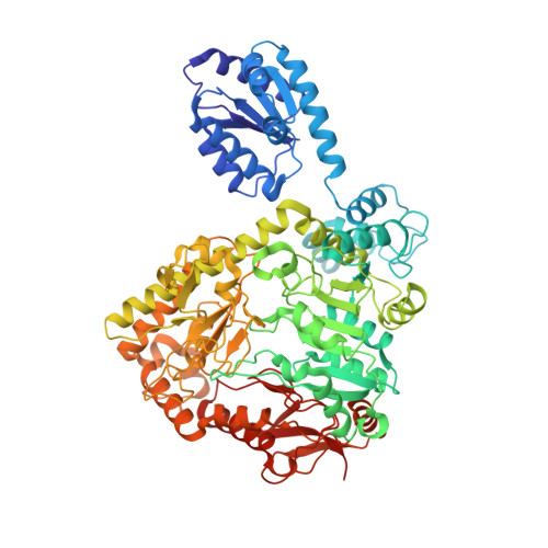
    The acid-induced arginine decarboxylase is part of an enzymatic system in Escherichia coli that contributes to making this organism acid resistant. The arginine decarboxylase is a vitamin B(6)-dependent enzyme that is active at acidic pH. It consumes a proton in the decarboxylation of arginine to agmatine, and by working in tandem with an arginine-agmatine antiporter, this enzymatic cycle protects the organism by preventing the accumulation of protons inside the cell. We have determined the structure of the acid-induced arginine decarboxylase by X-ray crystallography to 2.4 A resolution. The arginine decarboxylase structure revealed a ca. 800 kDa decamer composed as a pentamer of five homodimers. Each homodimer has an abundance of acidic surface residues, which at neutral pH prevents inactive homodimers from associating into active decamers. Conversely, acidic conditions favor the assembly of active decamers. Therefore, the structure of arginine decarboxylase presents a mechanism by which its activity is modulated by external pH.


  • Organizational Affiliation

    Division of Molecular Biosciences, Imperial College, London SW7 2AZ, UK.


Macromolecules
Find similar proteins by:  (by identity cutoff)  |  3D Structure
Entity ID: 1
MoleculeChains Sequence LengthOrganismDetailsImage
BIODEGRADATIVE ARGININE DECARBOXYLASE
A, B, C, D, E
A, B, C, D, E, F, G, H, I, J
755Escherichia coli BL21(DE3)Mutation(s): 0 
EC: 4.1.1.19
UniProt
Find proteins for P28629 (Escherichia coli (strain K12))
Explore P28629 
Go to UniProtKB:  P28629
Entity Groups  
Sequence Clusters30% Identity50% Identity70% Identity90% Identity95% Identity100% Identity
UniProt GroupP28629
Sequence Annotations
Expand
  • Reference Sequence
Experimental Data & Validation

Experimental Data

  • Method: X-RAY DIFFRACTION
  • Resolution: 2.40 Å
  • R-Value Free: 0.229 
  • R-Value Work: 0.177 
  • R-Value Observed: 0.179 
  • Space Group: P 64
Unit Cell:
Length ( Å )Angle ( ˚ )
a = 197.651α = 90
b = 197.651β = 90
c = 450.322γ = 120
Software Package:
Software NamePurpose
REFMACrefinement
DENZOdata reduction
SCALEPACKdata scaling
SHARPphasing

Structure Validation

View Full Validation Report


Ligand Structure Quality Assessment 


Entry History 

Deposition Data

Revision History  (Full details and data files)

  • Version 1.0: 2009-03-31
    Type: Initial release
  • Version 1.1: 2011-07-13
    Changes: Advisory, Version format compliance
  • Version 1.2: 2011-10-12
    Changes: Database references, Derived calculations, Other, Structure summary
  • Version 1.3: 2015-12-02
    Changes: Atomic model, Data collection, Derived calculations, Non-polymer description, Other, Refinement description, Structure summary