2VUO

Crystal structure of the rabbit IgG Fc fragment


Experimental Data Snapshot

  • Method: X-RAY DIFFRACTION
  • Resolution: 1.95 Å
  • R-Value Free: 0.204 
  • R-Value Work: 0.169 
  • R-Value Observed: 0.168 

wwPDB Validation   3D Report Full Report


This is version 2.1 of the entry. See complete history


Literature

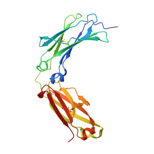
The Crystal Structure of Rabbit Igg-Fc.

Girardi, E.Holdom, M.D.Davies, A.M.Sutton, B.J.Beavil, A.J.

(2009) Biochem J 417: 77

  • DOI: https://doi.org/10.1042/BJ20081355
  • Primary Citation of Related Structures:  
    2VUO

  • PubMed Abstract: 

    We report the structure of the Fc fragment of rabbit IgG at 1.95 A (1 A=0.1 nm) resolution. Rabbit IgG was the molecule for which Porter established the four-chain, Upsilon-shaped structure of the antibody molecule, and crystals of the Fc ('Fragment crystallisable') were first reported almost 50 years ago in this journal [Porter, R. R. (1959) Biochem. J. 73, 119-126]. This high-resolution analysis, apparently of the same crystal form, reveals several features of IgG-Fc structure that have not previously been described. More of the lower hinge region is visible in this structure than in others, demonstrating not only the acute bend in the IgG molecule that this region can mediate, as seen in receptor complexes, but also that this region has a tendency to adopt a bent structure even in the absence of receptor. As observed in other IgG-Fc structures, the Cgamma2 domains display greater mobility/disorder within the crystals than the Cgamma3 domains; unexpectedly the structure reveals partial cleavage of both Cgamma2 intra-domain disulphide bonds, whereas an alternative conformation for one of the cysteine residues in the intact bridge within the more ordered Cgamma3 domains is observed. The N-linked oligosaccharide chains at Asn(297) are well-defined and reveal two alternative conformations for the galactose units on each of the alpha(1-6)-linked branches. The presence of this galactose unit is important for stabilizing the structure of the entire branched carbohydrate chain, and its absence correlates with the severity of autoimmune conditions such as rheumatoid arthritis in both human clinical studies and in a rabbit model of the disease. Rabbit IgG, through this high-resolution structure of its Fc region, thus continues to offer new insights into antibody structure.


  • Organizational Affiliation

    Randall Division of Cell and Molecular Biophysics, King's College London, New Hunt's House, Guy's Campus, London SE11UL, UK.


Macromolecules
Find similar proteins by:  (by identity cutoff)  |  3D Structure
Entity ID: 1
MoleculeChains Sequence LengthOrganismDetailsImage
IG GAMMA CHAIN C REGION
A, B
219Oryctolagus cuniculusMutation(s): 0 
UniProt
Find proteins for P01870 (Oryctolagus cuniculus)
Explore P01870 
Go to UniProtKB:  P01870
Entity Groups  
Sequence Clusters30% Identity50% Identity70% Identity90% Identity95% Identity100% Identity
UniProt GroupP01870
Sequence Annotations
Expand
  • Reference Sequence
Oligosaccharides

Help

Entity ID: 2
MoleculeChains Length2D Diagram Glycosylation3D Interactions
beta-D-galactopyranose-(1-4)-2-acetamido-2-deoxy-beta-D-glucopyranose-(1-2)-alpha-D-mannopyranose-(1-6)-[2-acetamido-2-deoxy-beta-D-glucopyranose-(1-2)-alpha-D-mannopyranose-(1-3)]beta-D-mannopyranose-(1-4)-2-acetamido-2-deoxy-beta-D-glucopyranose-(1-4)-2-acetamido-2-deoxy-beta-D-glucopyranose
C
8N-Glycosylation
Glycosylation Resources
GlyTouCan:  G89993FE
GlyCosmos:  G89993FE
GlyGen:  G89993FE
Entity ID: 3
MoleculeChains Length2D Diagram Glycosylation3D Interactions
beta-D-galactopyranose-(1-4)-2-acetamido-2-deoxy-beta-D-glucopyranose-(1-2)-alpha-D-mannopyranose-(1-6)-[alpha-D-mannopyranose-(1-3)]beta-D-mannopyranose-(1-4)-2-acetamido-2-deoxy-beta-D-glucopyranose-(1-4)-2-acetamido-2-deoxy-beta-D-glucopyranose
D
7N-Glycosylation
Glycosylation Resources
GlyTouCan:  G28663KH
GlyCosmos:  G28663KH
GlyGen:  G28663KH
Small Molecules
Ligands 3 Unique
IDChains Name / Formula / InChI Key2D Diagram3D Interactions
GOL
Query on GOL

Download Ideal Coordinates CCD File 
E [auth A],
F [auth A],
G [auth A],
K [auth B]
GLYCEROL
C3 H8 O3
PEDCQBHIVMGVHV-UHFFFAOYSA-N
FMT
Query on FMT

Download Ideal Coordinates CCD File 
I [auth A]
J [auth A]
M [auth B]
N [auth B]
O [auth B]
I [auth A],
J [auth A],
M [auth B],
N [auth B],
O [auth B],
P [auth B]
FORMIC ACID
C H2 O2
BDAGIHXWWSANSR-UHFFFAOYSA-N
AZI
Query on AZI

Download Ideal Coordinates CCD File 
H [auth A],
L [auth B]
AZIDE ION
N3
IVRMZWNICZWHMI-UHFFFAOYSA-N
Experimental Data & Validation

Experimental Data

  • Method: X-RAY DIFFRACTION
  • Resolution: 1.95 Å
  • R-Value Free: 0.204 
  • R-Value Work: 0.169 
  • R-Value Observed: 0.168 
  • Space Group: P 1 21 1
Unit Cell:
Length ( Å )Angle ( ˚ )
a = 58.662α = 90
b = 71.211β = 104.78
c = 69.036γ = 90
Software Package:
Software NamePurpose
PHENIXrefinement
MOSFLMdata reduction
SCALAdata scaling
MOLREPphasing

Structure Validation

View Full Validation Report


Entry History 

Deposition Data

Revision History  (Full details and data files)

  • Version 1.0: 2008-09-16
    Type: Initial release
  • Version 1.1: 2011-05-08
    Changes: Version format compliance
  • Version 1.2: 2011-07-13
    Changes: Version format compliance
  • Version 1.3: 2019-05-08
    Changes: Data collection, Derived calculations, Experimental preparation, Other
  • Version 1.4: 2019-05-22
    Changes: Data collection, Experimental preparation
  • Version 2.0: 2020-07-29
    Type: Remediation
    Reason: Carbohydrate remediation
    Changes: Atomic model, Data collection, Derived calculations, Other, Structure summary
  • Version 2.1: 2023-12-13
    Changes: Data collection, Database references, Derived calculations, Refinement description, Structure summary