2VPV

Dimerization Domain of Mif2p


Experimental Data Snapshot

  • Method: X-RAY DIFFRACTION
  • Resolution: 2.70 Å
  • R-Value Free: 0.242 
  • R-Value Work: 0.208 
  • R-Value Observed: 0.210 

wwPDB Validation   3D Report Full Report


This is version 1.3 of the entry. See complete history


Literature

Structural and Functional Dissection of Mif2P, a Conserved DNA-Binding Kinetochore Protein.

Cohen, R.L.Espelin, C.W.De Wulf, P.Sorger, P.K.Harrison, S.C.Simons, K.T.

(2008) Mol Biol Cell 19: 4480

  • DOI: https://doi.org/10.1091/mbc.e08-03-0297
  • Primary Citation of Related Structures:  
    2VPV

  • PubMed Abstract: 

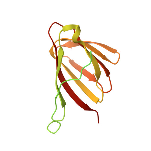
    Mif2p is the budding-yeast orthologue of the mammalian centromere-binding protein CENP-C. We have mapped domains of Saccharomyces cerevisiae Mif2p and studied the phenotyptic consequences of their deletion. Using chromatin immunoprecipitation (ChIP) and electrophoretic mobility shift assays, we have further shown that Mif2p binds in the CDEIII region of the budding-yeast centromere, probably in close spatial association with Ndc10p. Moreover, ChIP experiments show that Mif2p recruits to yeast kinetochores a substantial subset of inner and outer kinetochore proteins, but not the Ndc80 or Spc105 complexes. We have determined the crystal structure of the C-terminal, dimerization domain of Mif2p. It has a "cupin" fold, extremely similar both in polypeptide chain conformation and in dimer geometry to the dimerization domain of a bacterial transcription factor. The Mif2p dimer seems to be part of an enhanceosome-like structure that nucleates kinetochore assembly in budding yeast.


  • Organizational Affiliation

    Jack and Eileen Connors Structural Biology Laboratory, Harvard Medical School, Boston, MA 02115, USA.


Macromolecules
Find similar proteins by:  (by identity cutoff)  |  3D Structure
Entity ID: 1
MoleculeChains Sequence LengthOrganismDetailsImage
PROTEIN MIF2
A, B
166Saccharomyces cerevisiaeMutation(s): 0 
UniProt
Find proteins for P35201 (Saccharomyces cerevisiae (strain ATCC 204508 / S288c))
Explore P35201 
Go to UniProtKB:  P35201
Entity Groups  
Sequence Clusters30% Identity50% Identity70% Identity90% Identity95% Identity100% Identity
UniProt GroupP35201
Sequence Annotations
Expand
  • Reference Sequence
Small Molecules
Ligands 1 Unique
IDChains Name / Formula / InChI Key2D Diagram3D Interactions
SO4
Query on SO4

Download Ideal Coordinates CCD File 
C [auth A]SULFATE ION
O4 S
QAOWNCQODCNURD-UHFFFAOYSA-L
Experimental Data & Validation

Experimental Data

  • Method: X-RAY DIFFRACTION
  • Resolution: 2.70 Å
  • R-Value Free: 0.242 
  • R-Value Work: 0.208 
  • R-Value Observed: 0.210 
  • Space Group: P 21 3
Unit Cell:
Length ( Å )Angle ( ˚ )
a = 106.02α = 90
b = 106.02β = 90
c = 106.02γ = 90
Software Package:
Software NamePurpose
REFMACrefinement
SOLVEphasing

Structure Validation

View Full Validation Report


Entry History 

Deposition Data

Revision History  (Full details and data files)

  • Version 1.0: 2008-08-26
    Type: Initial release
  • Version 1.1: 2011-06-02
    Changes: Version format compliance
  • Version 1.2: 2011-07-13
    Changes: Version format compliance
  • Version 1.3: 2019-05-08
    Changes: Advisory, Data collection, Derived calculations, Experimental preparation, Other