2VM0

Crystal structure of radiation-induced myoglobin compound II generated after annealing of peroxymyoglobin


Experimental Data Snapshot

  • Method: X-RAY DIFFRACTION
  • Resolution: 1.60 Å
  • R-Value Free: 0.190 
  • R-Value Work: 0.163 
  • R-Value Observed: 0.165 

wwPDB Validation   3D Report Full Report


This is version 1.3 of the entry. See complete history


Literature

The Crystal Structure of Peroxymyoglobin Generated Through Cryoradiolytic Reduction of Myoglobin Compound III During Data Collection.

Hersleth, H.-P.Hsiao, Y.Ryde, U.Gorbitz, C.H.Andersson, K.K.

(2008) Biochem J 412: 257

  • DOI: https://doi.org/10.1042/BJ20070921
  • Primary Citation of Related Structures:  
    2VLX, 2VLY, 2VLZ, 2VM0

  • PubMed Abstract: 

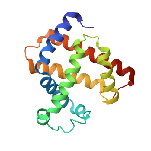
    Myoglobin has the ability to react with hydrogen peroxide, generating high-valent complexes similar to peroxidases (compounds I and II), and in the presence of excess hydrogen peroxide a third intermediate, compound III, with an oxymyoglobin-type structure is generated from compound II. The compound III is, however, easily one-electron reduced to peroxymyoglobin by synchrotron radiation during crystallographic data collection. We have generated and solved the 1.30 A (1 A=0.1 nm) resolution crystal structure of the peroxymyoglobin intermediate, which is isoelectric to compound 0 and has a Fe-O distance of 1.8 A and O-O bond of 1.3 A in accordance with a Fe(II)-O-O- (or Fe(III)-O-O2-) structure. The generation of the peroxy intermediate through reduction of compound III by X-rays shows the importance of using single-crystal microspectrophotometry when doing crystallography on metalloproteins. After having collected crystallographic data on a peroxy-generated myoglobin crystal, we were able (by a short annealing) to break the O-O bond leading to formation of compound II. These results indicate that the cryoradiolytic-generated peroxymyoglobin is biologically relevant through its conversion into compound II upon heating. Additionally, we have observed that the Xe1 site is occupied by a water molecule, which might be the leaving group in the compound II to compound III reaction.


  • Organizational Affiliation

    University of Oslo, Department of Molecular Biosciences, P.O. Box 1041 Blindern, N-0316 Oslo, Norway.


Macromolecules
Find similar proteins by:  (by identity cutoff)  |  3D Structure
Entity ID: 1
MoleculeChains Sequence LengthOrganismDetailsImage
MYOGLOBIN153Equus caballusMutation(s): 0 
UniProt
Find proteins for P68082 (Equus caballus)
Explore P68082 
Go to UniProtKB:  P68082
Entity Groups  
Sequence Clusters30% Identity50% Identity70% Identity90% Identity95% Identity100% Identity
UniProt GroupP68082
Sequence Annotations
Expand
  • Reference Sequence
Small Molecules
Ligands 5 Unique
IDChains Name / Formula / InChI Key2D Diagram3D Interactions
HEM
Query on HEM

Download Ideal Coordinates CCD File 
B [auth A]PROTOPORPHYRIN IX CONTAINING FE
C34 H32 Fe N4 O4
KABFMIBPWCXCRK-RGGAHWMASA-L
SO4
Query on SO4

Download Ideal Coordinates CCD File 
D [auth A],
E [auth A]
SULFATE ION
O4 S
QAOWNCQODCNURD-UHFFFAOYSA-L
GOL
Query on GOL

Download Ideal Coordinates CCD File 
F [auth A]GLYCEROL
C3 H8 O3
PEDCQBHIVMGVHV-UHFFFAOYSA-N
PEO
Query on PEO

Download Ideal Coordinates CCD File 
G [auth A],
H [auth A],
I [auth A]
HYDROGEN PEROXIDE
H2 O2
MHAJPDPJQMAIIY-UHFFFAOYSA-N
OH
Query on OH

Download Ideal Coordinates CCD File 
C [auth A]HYDROXIDE ION
H O
XLYOFNOQVPJJNP-UHFFFAOYSA-M
Experimental Data & Validation

Experimental Data

  • Method: X-RAY DIFFRACTION
  • Resolution: 1.60 Å
  • R-Value Free: 0.190 
  • R-Value Work: 0.163 
  • R-Value Observed: 0.165 
  • Space Group: P 1 21 1
Unit Cell:
Length ( Å )Angle ( ˚ )
a = 63.723α = 90
b = 28.73β = 105.78
c = 35.383γ = 90
Software Package:
Software NamePurpose
REFMACrefinement
MOSFLMdata reduction
SCALAdata scaling
CNSphasing

Structure Validation

View Full Validation Report


Entry History 

Deposition Data

Revision History  (Full details and data files)

  • Version 1.0: 2008-01-29
    Type: Initial release
  • Version 1.1: 2011-06-02
    Changes: Version format compliance
  • Version 1.2: 2011-07-13
    Changes: Version format compliance
  • Version 1.3: 2023-12-13
    Changes: Data collection, Database references, Derived calculations, Other, Refinement description